Encephalitozoonosis
The Encephalitozoonose ( "stargazers disease") is a single-celled by the Encephalitozoon cuniculi , rare Encephalitozoon intestinalis or Encephalitozoon light, caused parasitic disease that in Europe especially rabbit attacks. Other strains of the pathogen cause disease in old world mice and canines . Encephalitozoonosis occurs mainly in immunocompromised animals. It is a potential zoonosis and, albeit very rarely, can also occur in immunocompromised people. The disease was first described by Wright and Craighead in 1922 .
The pathogen mainly affects the kidneys and the brain . The latter manifests itself in neurological disorders, with tilted head being the most common symptom . The antiparasitic agent fenbendazole can be used to combat the pathogen and thus new infections. Upon the occurrence of clinical symptoms that needs treatment by administration of antibiotics to be extended and supportive measures that cure view is then uncertain.
Pathogen and occurrence
Encephalitozoon cuniculi is a single cell from the group of microsporidia that onlylivesin cells of higher organisms ( obligate intracellularly ). Like all microsporidia, it is an organism closely related to fungi with a cell nucleus and cell membrane ( eukaryote ), but whichlackssome cell organelles such as mitochondria . The genome isextremely smallwith 2.9 million base pairs thatencodejust under 2000 proteins . In mammals, the parasite attacks the cells of the kidney, brain and other organs. Outside of its host , the unicellular organism survives in the form of a 2 µm spore , which represents the infectious permanent stage.
Depending on the main host, three different strains of Encephalitozoon cuniculi are distinguished. In principle, rabbits are susceptible to all three, but natural infections have so far only been described for the rabbit strain. The following tribes occur:
- In Europe, the rabbit strain (type I), which occurs worldwide, plays a major role. Previous studies found antibodies in 7 to 52% of domestic rabbits in healthy animals . However, this seroprevalence only shows that the animals had contact with the pathogen and that there is a high probability that they still carry it. A disease only occurs with a temporary disturbance of the immune system , e.g. B. after viral infections. The seroprevalence of neurologically diseased domestic rabbits is up to 85%. The pathogen reservoir is presumably made up of wild rabbits , in which the seroprevalence is between 4 and 25%; other rabbits are apparently not carriers of the pathogen. Encephalitozoonosis is now the most common infectious disease in domestic rabbits.
- Encephalitozoon cuniculi type II (strain of mice) is primarily a disease-causing agent for old -world mice and has so far only been detected in Europe. The seroprevalence in rats and mice living in the wild is between 3 and 4%; the pathogen is practically non-existent in laboratory facilities due to the high hygiene standards. Fatal infections with this type have also been observed in farm foxes in Scandinavia .
- Encephalitozoon cuniculi type III (dog strain) is widespread in North America and South Africa, mainly affects dogs and is probably the only potentially disease-causing ( pathogenic ) strain for them. Infections in monkeys have also been observed in zoos around the world .
E. cuniculi occurs worldwide; the disease was first described in rabbits in 1922. Antibodies against E. cuniculi can be detected in many mammals . Reports of human disease are limited to immunocompromised and AIDS patients, with only the rabbit and dog strains believed to be potentially dangerous. In eastern Slovakia the seroprevalence was 5.7%, in people with immunodeficiencies even 37.5%. The seroprevalence in horses is between 14% and 60%.
Route of infection and disease development
The most common type of transmission appears to be oral ingestion of the spores, which are mainly excreted in the urine. A transmission of the pathogen from the mother to the fetuses before birth ( intrauterine ) is also possible. After the spores have been absorbed, the pathogen is absorbed by scavenger cells ( phagocytes ) in the intestine and distributed with them through the bloodstream.
The infection does not usually cause disease. The host reacts to invasion of the pathogen with an immune reaction that is mediated by cytotoxic CD8 (+) T cells .
An outbreak of disease may not occur until years after infection if the immune system is disrupted , for example when the animals are exposed to noise and stress. In rabbits, the pathogen mainly colonizes the kidneys , where it causes chronic kidney inflammation with proliferation or atrophy of the epithelium of the renal tubules . In the brain and meninges , purulent inflammation ( meningoencephalitis ) with proliferation ( gliosis ) of astrocytes and lymphocyte infiltration around the blood vessels does not occur until chronic infection . In addition, spores can settle in the lens of the eye and trigger phacoclastic uveitis , but this localization seems to only occur if it is transmitted in the womb. In tamarins , inflammation of the heart muscle , liver , lungs , skeletal muscle and retina was also found . In immunosuppressed mice, non-purulent, lymphocytic meningoencephalitis with destruction of nerve cells and astrogliosis was found. Horses can a necrotizing inflammation of the placenta ( placentitis develop).
Symptoms
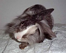
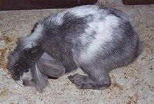
The classic symptoms of Encephalitozoonose in rabbits are neurological disorders such as torticollis ( torticollis ), usually in combination with nystagmus ( nystagmus ), impaired locomotor coordination ( ataxia ), stiff gait, paralysis and convulsions. In the advanced course of the disease, animals with severe brain affections often turn uncontrollably around their own longitudinal axis and can be seriously injured in the process. The disease can also manifest itself in the form of renal insufficiency or lens opacity and inflammation of the middle skin of the eye after rupture of the lens capsule ( phacoclastic uveitis ). In one study, 45% of the infected rabbits showed neurological deficits, 31% kidney symptoms and 14% uveitis. Especially in the case of outdoor posture, there is a risk of fly maggot infestation in the case of neurological disorders due to the restricted mobility and thus personal hygiene .
In dogs and foxes, encephalitozoonosis manifests itself in kidney failure and central nervous symptoms that resemble distemper . Such diseases have so far only been observed in dogs in Africa and the United States, while diseases in foxes have also occurred in Scandinavia. In cats, eye infections (phacoclastic uveitis, focal lens opacity , anterior uveitis) occur primarily, with the mouse strain (type II) being the main trigger.
In other animals the symptoms of the disease are mostly unspecific and encephalitozoonosis is only discovered during pathological dissection. Stillbirths and sudden juvenile deaths occur in semi-monkeys. The importance of serological detection in horses has not yet been clarified: Encephalitozoon cuniculi can cause abortions, but is also discussed in connection with colic and neurological disorders. Symptoms in people who are immunocompromised or infected with HIV are described in the “ Danger to Humans ” section.
Diagnosis
The diagnosis cannot be made with certainty in living animals.
The clinical diagnosis is always a suspected diagnosis. Since many domestic rabbits carry the pathogen without getting sick, a serological test for antibodies (India-Ink immunoreaction, titer determination by indirect immunofluorescence ) against the pathogen gives an indication of an infection, but whether the existing symptoms are caused by it , must be clarified by excluding other diseases. An antibody titer can also be detected in over 40% of healthy rabbits. A study found mean titers of 1: 1324 in rabbits with clinical suspicion, which is around 1.7 times higher than in animals without one. In contrast, further studies could not prove a connection between the level of the titre and the disease. In addition, antibody levels can remain high for years after infection. Rabbits that have already become infected in the womb usually do not show any antibodies since the pathogen is not recognized as foreign (→ self-tolerance ). Direct detection of the pathogen DNA by means of PCR in urine, feces or cerebral fluid is rarely successful. In addition, pathogen DNA does not appear in the urine until three to five weeks after infection and also in some healthy animals. Only in the case of phacoclastic uveitis can the diagnosis be made unequivocally using PCR on removed lens material.
The main symptom "torticollis" can also occur in rabbits with inflammation of the inner ear ( otitis interna , main pathogen Pasteurella multocida ), viral infections of the brain, listeriosis , toxoplasmosis , wandering larvae ( larva migrans ) of raccoon roundworm , tumors (especially lymphomas ) and abscesses of the brain as well as head injuries occur. Cardiovascular diseases, poisoning, metabolic disorders or spinal cord trauma can also cause neurological failure symptoms. Some of these diseases can be detected by imaging methods and thus indirectly exclude encephalitozoonosis.
A reliable diagnosis is only possible after death by a pathological examination with detection of the pathogen. It can be detected by immunohistochemistry or PCR. Cultivation is possible, but very complex.
Danger to humans
Encephalitozoonosis is a potential zoonosis , but so far only diseases have been observed in people with a severe weakening of the immune system (e.g. AIDS patients, people with immunosuppression after organ transplants, idiopathic CD4 + T lymphocytopenia ). Theoretically, people with a weak immune system (young children, very old people) could also be receptive, but there is no evidence yet.
In most cases, sick animals have already excreted the pathogens over a long period of time. Although a large number of pet rabbits are seropositive, there is so far no evidence that a human has been infected from a rabbit or other animal, although the route of infection in humans has not yet been clarified. There is one case of human-to-human transmission of the canine strain in a bone marrow transplant in a Hodgkin's disease patient who subsequently died of pneumonia .
In people with immunodeficiency, however, diarrheal diseases as a result of infections with Encephalitozoon bieneusi and Encephalitozoon intestinalis play a far greater role, while Encephalitozoon cuniculi infections are very rare even in this group of people. The symptoms of such a disease range from fever , chest, stomach, muscle and headache, cough, runny nose, diarrhea, sinus and pneumonia, conjunctivitis and corneal inflammation to kidney failure . Encephalitozoon brightem can also cause both keratoconjunctivis and disseminated infections in humans. Since 1994, only 17 E. cuniculi infections have been detected in AIDS patients and 6 in people after organ transplants worldwide. Older case descriptions must be interpreted with caution, since encephalitozoon species cannot be differentiated by light microscopy and the molecular biological detection methods were only established in the 1990s.
treatment
There is currently no 100 percent effective treatment for encephalitozoonosis.
Eliminating the pathogen in rabbits is probably not possible, because quite a few animals that improve clinically with treatment are presented at a later point in time with repeated symptoms. The antiparasitic drugs fenbendazole and albendazole only lead to a reduction in the number of pathogens and can limit new infections; however, the effect is limited in the event of a clinical outbreak of an encephalitozoon cuniculi infection. Since the rabbits are immunocompromised at the time of the onset of the disease, the administration of an antibiotic ( chloramphenicol , gyrase inhibitor , chloroquine phosphate , oxytetracycline or sulfonamides ) is recommended. Glucocorticoids are also used to reduce inflammation , but their use is controversial, as they can also suppress the body's own T-cell response and often cause severe side effects in rabbits. In addition, the animals should be given infusions, especially if they have renal insufficiency. This requires regular blood tests. The administration of a vitamin B complex is also recommended by some authors as a supportive measure. Rabbits with symptoms of paralysis should receive additional physiotherapy treatment by moving the paralyzed limbs. Rabbits that do not eat their own food must be force-fed. Noise and stress are to be kept away from the sick animal in any case. It should be remembered that animals have a different hearing threshold than humans and thus perceive noises that cannot be recognized by humans. In the case of a disease of the eye, only removal of the lens protein that has leaked from the ruptured lens capsule can lead to a cure. If this does not happen, episodes with severe uveitides will occur again and again.
In immunosuppressed people with encephalitozoonosis, albendazole is used against encephalitozoon cuniculi and other microsporidia. Newer therapeutic approaches are polyamines , chitin inhibitors such as nikkomycin and fluoroquinolones , and fumagillin for local eye inflammation .
The potential risk of animal-to-human transmission can be minimized through consistent hygiene measures. In addition to the daily removal of faeces and urine, this includes cleaning the cage or enclosure with cleaning and disinfecting agents. For disinfection is boiling water are suitable, 2-percent Lysol , 1-percent formaldehyde or 70-percent alcohol . After contact with animals, hands should be washed thoroughly, also to reduce the risk of transmission of other zoonoses.
Healing prospects
In some cases, rabbits heal spontaneously without therapy. Clinical healing of inclined head posture and ataxias is, however, generally more beneficial the faster the therapy is started. If the neurological symptoms have been present for a long time, a significantly longer time must be expected for complete healing ( restitutio ad integrum ). Sometimes, in particularly severe cases, it can take several months after the drug treatment is completed for the head tilt to go away. However, the disease can also lead to permanent damage to the brain, resulting in permanent head tilt. A relapse is always to be expected, but preventive, long-term administration of fenbendazole is not recommended, as the pathogen can develop resistance to the active substance and the substance can also have an immunosuppressive effect. Serious infections can also be fatal or cause such permanent impairment that euthanasia is indicated.
literature
- Peter Deplazes: Encephalitozoonosis. In: Andre Jaggy: Atlas and textbook of small animal neurology. Schlütersche 2005, ISBN 3-87706-739-5 , p. 458.
- Anja Ewringmann: Key symptoms in rabbits. Diagnostic guide and therapy. Enke-Verlag, 2004, ISBN 3-8304-1020-4 .
- EJ Gentz and JW Carpenter: Neurologic and musculoskeletal diseases. In: EV Hillyer and KE Quesenberry (Eds.): Ferrets, rabbits, and rodents. Saunders 1999, ISBN 0-7216-4023-0 , pp. 220-226.
- Frances Harcourt-Brown: Textbook of rabbit medicine. Butterworth-Heinemann, 2004, ISBN 0-7506-4002-2 .
- Thomas Schnieder (Ed.): Veterinary Parasitology. Paul Parey, 2006, ISBN 3-8304-4135-5 .
- Ulrike Flock: Encephalitozoonosis in rabbits - a retrospective evaluation. (PDF; 606 kB), Dissertation, Veterinary Faculty of the Ludwig-Maximilians-Universität, Munich 2010, accessed September 17, 2013.
Web links
- B. Drescher: Encephalitozoonosis in rabbits
- Intervet: encephalitozoonosis
Individual evidence
- ↑ a b Thomas Göbel: Encephalitozoonosis: Most common infectious disease in rabbits . In: VETImpulse. 15, issue 18, 2006.
- ↑ ES Didier, CR Vossbrinck, MD Baker, LB Rogers, DC Bertucci, YES Shadduck: Identification and characterization of three Encephalitozoon cuniculi strains . In: Parasitology . 111 (Pt 4), November 1995, ISSN 0031-1820 , p. 411-421 , PMID 11023405 .
- ^ A. Mathis, M. Michel, H. Kuster, C. Müller, R. Weber, P. Deplazes: Two Encephalitozoon cuniculi strains of human origin are infectious to rabbits . In: Parasitology . 114 (Pt 1), January 1997, ISSN 0031-1820 , p. 29-35 , PMID 9011071 .
- ↑ a b c d e Frank Künzel, Anja Joachim: Encephalitozoonosis in rabbits . In: Parasitol. Res. Band 106 , 2010, p. 299-309 , doi : 10.1007 / s00436-009-1679-3 .
- ↑ EJ Keeble, DJ Shaw: Seroprevalence of antibodies to Encephalitozoon cuniculi in domestic rabbits in the United Kingdom . In: The Veterinary Record . tape 158 , no. 16 , April 22, 2006, ISSN 0042-4900 , p. 539-544 , PMID 16632526 .
- ↑ a b c d e f g Alexander Mathis, Rainer Weber, Peter Deplazes: Zoonotic potential of the microsporidia . In: Clinical Microbiology Reviews . tape 18 , no. 3 , 2005, ISSN 0893-8512 , p. 423-445 , doi : 10.1128 / CMR.18.3.423-445.2005 , PMID 16020683 , PMC 1195965 (free full text).
- ↑ a b C. Juan-Sallés, MM Garner, ES Didier, S. Serrato, LD Acevedo, JA Ramos-Vara, RW Nordhausen, LC Bowers, A. Parás: Disseminated encephalitozoonosis in captive, juvenile, cotton-top (Saguinus oedipus ) and neonatal emperor (Saguinus imperator) tamarins in North America . In: Veterinary Pathology . tape 43 , no. 4 , July 2006, ISSN 0300-9858 , p. 438-446 , doi : 10.1354 / vp.43-4-438 , PMID 16846985 .
- ↑ Monika Halánová, Lýdia Cisláková, Alexandra Valencákova, Pavol Bálent, Jozef Adam, Milan Trávnicek: Serological screening of occurrence of antibodies to Encephalitozoon cuniculi in humans and animals in Eastern Slovakia . In: Annals of agricultural and environmental medicine: AAEM . tape 10 , no. 1 , 2003, ISSN 1232-1966 , p. 117-120 , PMID 12852743 .
- ^ David Goodwin, Solange M. Gennari, Daniel K. Howe, JP Dubey, Anne M. Zajac, David S. Lindsay: Prevalence of antibodies to Encephalitozoon cuniculi in horses from Brazil . In: Veterinary Parasitology . tape 142 , no. 3-4 , December 20, 2006, ISSN 0304-4017 , p. 380–382 , doi : 10.1016 / j.vetpar.2006.07.006 , PMID 16919878 .
- ↑ a b Mária Levkutová, Vlasta Hípiková, Shay Faitelzon, Gad Benath, Stefan Paulík, Mikulás Levkut: Prevalence of antibodies to Encephalitozoon cuniculi in horses in the Israel . In: Annals of agricultural and environmental medicine: AAEM . tape 11 , no. 2 , 2004, ISSN 1232-1966 , p. 265-267 , PMID 15627335 .
- ↑ PJR Baneux, F. Pognan: In utero transmission of Encephalitozoon cuniculi strain type I in rabbits . In: Laboratory Animals . tape 37 , no. 2 , April 2003, ISSN 0023-6772 , p. 132-138 , doi : 10.1258 / 00236770360563778 , PMID 12689424 .
- ^ IA Khan, M. Moretto, LM Weiss: Immune response to Encephalitozoon cuniculi infection . In: Microbes and Infection / Institut Pasteur . tape 3 , no. 5 , 2001, ISSN 1286-4579 , p. 401-405 , PMC 3109655 (free full text).
- ↑ JC Cox, RC Hamilton, HD Attwood: An investigation of the route and progression of Encephalitozoon cuniculi infection in adult rabbits . In: The Journal of Protozoology . tape 26 , no. 2 , May 1979, ISSN 0022-3921 , pp. 260-265 , PMID 490434 .
- ↑ Maria Anete Lallo, Eduardo Fernandes Bondan: Experimental meningoencephalomyelitis by Encephalitozoon cuniculi in cyclophosphamide-immunosuppressed mice . In: Arquivos De Neuro-Psiquiatria . tape 63 , 2A, June 2005, ISSN 0004-282X , p. 246-251 , PMID 16100970 .
- ↑ a b J. C. Patterson-Kane, P. Caplazi, F. Rurangirwa, RR Tramontin, K. Wolfsdorf: Encephalitozoon cuniculi placentitis and abortion in a quarterhorse mare . In: Journal of Veterinary Diagnostic Investigation: Official Publication of the American Association of Veterinary Laboratory Diagnosticians, Inc . tape 15 , no. 1 , January 2003, ISSN 1040-6387 , p. 57-59 , PMID 12580298 .
- ↑ C. Giordano, A. Weigt, A. Vercelli, M. Rondena, G. Grilli, C. Giudice: Immunohistochemical identification of Encephalitozoon cuniculi in phacoclastic uveitis in four rabbits . In: Veterinary Ophthalmology . tape 8 , no. 4 , 2005, ISSN 1463-5216 , p. 271-275 , doi : 10.1111 / j.1463-5224.2005.00394.x , PMID 16008708 .
- ↑ Anja Ewringmann, Thomas Göbel: Investigations on the clinic and therapy of encephalitozoonosis in pet rabbits . In: Small Animal Practice . tape 44 , no. 5 , May 1999, pp. 357-372 .
- ^ WS Botha, AF van Dellen, CG Stewart: Canine encephalitozoonosis in South Africa . In: Journal of the South African Veterinary Association . tape 50 , no. 2 , June 1979, ISSN 1019-9128 , pp. 135-144 , PMID 551193 .
- ↑ Wilson Jm: Encephalitozoon cuniculi in wild European rabbits and a fox. In: Research in veterinary science . tape 26 , no. 1 , January 1979, p. 114 , PMID 472479 .
- ↑ J. Akerstedt, K. Nordstoga, A. Mathis, E. Smeds, P. Deplazes: Fox encephalitozoonosis: isolation of the agent from an outbreak in farmed blue foxes (Alopex lagopus) in Finland and some hitherto unreported pathologic lesions . In: Journal of Veterinary Medicine. B, Infectious Diseases and Veterinary Public Health . tape 49 , no. 8 , October 2002, ISSN 0931-1793 , p. 400-405 , PMID 12449250 .
- ↑ Petra Benz, Günter Maaß, Jacqueline Csokai, Andrea Fuchs-Baumgartinger, Ilse Schwendenwein, Alexander Tichy, Barbara Nell: Detection of Encephalitozoon cuniculi in the feline cataractous lens . In: Veterinary Ophthalmology . 14, Suppl. 1, September 2011, p. 37-47 , doi : 10.1111 / j.1463-5224.2011.00873.x .
- ↑ Carolyn Cray, Giselle Arcia, Renata Schneider, Susan A. Kelleher, Kristopher L. Arheart: Evaluation of the usefulness of an ELISA and protein electrophoresis in the diagnosis of Encephalitozoon cuniculi infection in rabbits . In: American Journal of Veterinary Research . tape 70 , no. 4 , March 31, 2009, p. 478-482 , doi : 10.2460 / ajvr.70.4.478 .
- ↑ a b c d Jaqueline Csokai: Head tilted position - encephalitozoonosis? What diagnostic options are there? In: Veterinary mirror . No. 2 , 2015, p. 72-75 , doi : 10.1055 / s-0034-1396194 .
- ↑ Frank Künzel, Andrea Gruber, Alexander Tichy, Renate Edelhofer, Barbara Nell, Jasmin Hassan, Michael Leschnik, Johann G. Thalhammer, Anja Joachim: Clinical symptoms and diagnosis of encephalitozoonosis in pet rabbits . In: Veterinary Parasitology . tape 151 , no. 2–4 , February 14, 2008, pp. 115–124 , doi : 10.1016 / j.vetpar.2007.11.005 .
- ↑ JM Orenstein, P. Russo, ES Didier, C. Bowers, N. Bunin, DT Teachey: Fatal pulmonary microsporidiosis due to encephalitozoon cuniculi following allogeneic bone marrow transplantation for acute myelogenous leukemia . In: Ultrastructural Pathology . tape 29 , no. 3-4 , 2005, ISSN 0191-3123 , p. 269-276 , doi : 10.1080 / 01913120590951257 , PMID 16036880 .
- ^ Marianne Abele-Horn: Antimicrobial Therapy. Decision support for the treatment and prophylaxis of infectious diseases. With the collaboration of Werner Heinz, Hartwig Klinker, Johann Schurz and August Stich, 2nd, revised and expanded edition. Peter Wiehl, Marburg 2009, ISBN 978-3-927219-14-4 , p. 292 f.
- ^ A b F. M. Harcourt-Brown, HKR Holloway: Encephalitozoon cuniculi in pet rabbits . In: The Veterinary Record . tape 152 , no. April 14 , 2003, ISSN 0042-4900 , p. 427-431 , PMID 12708591 .
- ↑ LM Felchle and RL Sigler: Phacoemulsification for the management of Encephalitozoon cuniculi-induced phacoclastic uveitis in a rabbit . In: Vet Ophthalmol . tape 5 , no. 3 , 2002, p. 211-215 , PMID 12236874 .
- ↑ ES Didier et al .: Therapeutic strategies for human microsporidia infections . In: Expert Rev Anti Infect Ther . tape 3 , no. 3 , 2005, p. 419-434 , PMID 15954858 .


