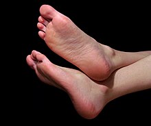
foot
The foot is the lowest section of the leg of the terrestrial vertebrate . In humans it consists of the tarsus , the metatarsus and the five free toes. Different types of feet are distinguished according to their external shape .
etymology
The common. Body part name mhd. Vuoz , ahd. Fuoz - for the Proto-Germanic. to be reconstructed is a root noun * fōt- with nom. Pl. * fōtiz , which in Ahd. has changed to the i -stems - is based on the ablaut form * pō̆d- of uridg. * pēd- "foot". Compare Latin pes , genitive pedis and altgr. πούς pous , genitive ποδός podos .
In Urgerm. was the Nom. Sg. originally probably * foz ; In the plural, the inflection as a consonant stem is still preserved in Old English, Old Frisian, and North Germanic. Compare also new Gl. feet , which ultimately goes to the Urm. Nom. Pl. * Fōtiz goes back; according to the law, this requires a form * pōdes and is therefore formally identical with urkelt. * ādes or better * ɸādes , documented in the Hesychgloss ἄδες · πόδες.
General
The foot consists of toes ( digiti pedis ), the middle foot ( metatarsus ) and a tarsus ( tarsus ). On the metatarsus, a distinction is made between the ball of the foot , the sole, the heel , the instep ( back of the foot) and the instep (outer edge). There are 26 bones in each of the feet (plus 2 sesamoid bones ), so the foot bones together make up about a quarter of the total of 206 to 215 bones in the human body .
The receptors of the skin senses ( sense of touch ) are found in a particularly high density on the soles of the feet and toes . The foot muscles have the task of executing the movements of the foot. In addition, it also tightens the longitudinal and transverse arches of the foot. The foot muscles are divided into the group of long and short foot muscles. The short foot muscles are located on the foot skeleton , which means that they have their origin and insertion here . The long foot muscles, on the other hand, lie on the lower leg .
Based on the difference in length between the big toe and the second toe, a distinction is made between three foot shapes:
- Egyptian foot: The second toe is shorter than the big toe.
- Greek foot: The second toe is longer than the big toe.
- Roman foot (also called square foot): The second toe and the big toe are the same length.
Even today, typical foot shapes can still be seen in people in certain regions and countries. There is also a certain variability, depending on the “use” of the foot: The Twa ( hunters and gatherers who often climb trees to harvest honey) and the Bakiga ( sedentary farmers) in Uganda have distinctively different feet.
Women have smaller feet than men relative to their body. It was also shown that small feet in women are perceived as attractive by both men and women, while large feet are perceived as masculine.
Evolutive development
→ See also upright gait in the article Hominization
The structure of the human foot and the hand made up of five rays are developments that occur for the first time in land vertebrates and are widely spread there. In individual groups of terrestrial vertebrates, the number of rays is reduced, for example in tailed amphibians, crocodiles and birds, and within mammals in cloven-hoofed and odd-toed ungulates.
Arch of the foot
The foot has a longitudinal arch and a transverse arch. As a result, the body weight is mainly carried by the three points of the heel, the big toe joint (ball of the big toe) and the big toe joint (ball of the small toe).
The arch of the foot (also known as the arch of the foot ) is tensed up by muscles and maintained by ligaments. The interaction of the tibialis posterior and the peroneus longus muscles is particularly important for maintaining the transverse vault . In addition, the transverse tracts of the plantar aponeurosis are important for the transverse vault as well as the transverse head of the adductor hallucis muscle . The sole tendon plate (aponeurosis plantaris) and the long sole ligament ( ligamentum plantare longum ) are important for maintaining the longitudinal arch . The longitudinal arch is tensed by the flexor hallucis longus and flexor digitorum longus muscles and the short muscles of the foot.
The arches of the foot are very important for the proper functioning of the foot, as they act like shock absorbers. Some diseases of the foot such as flat feet , arched feet and splayfoot are based on a sagging of the arch of the foot. An excessively pronounced arch of the foot also impairs the function of the foot, which is referred to as a hollow foot .
Sole of the foot
The sole of the foot ( Planta pedis or Planta for short ) has a substructure made of a body of fat that absorbs shocks and has a cushioning effect, but is so stable that it cannot slip under the forces acting when walking. Anatomical features can hardly be felt through this fat body, with the exception of the metatarsal heads of the central rays.
The sole of the foot can be divided into the following areas, which can be seen in a footprint in the sand or in the footprint of a doctor:
- heel
- Outer edge of the foot
- Area of the longitudinal vault
- Ball of the foot with the balls of the big toe and the ball of the little toe under the metatarsophalangeal joint of the big toe and the metatarsophalangeal of the little toe and all other toe balls under the other metatarsophalangean joints
The sole of the foot is not in contact with the ground. In the area of the longitudinal arch or the inner edge of the foot, it does not rest on a healthy foot. The body weight is borne by the sole of the foot in different proportions. The main part of the body weight is carried by the heel (approx. 33%) and the ball of the foot (approx. 40%). The rest is done by the outer edge of the foot (approx. 15%), the big toe (approx. 5%) and the remaining toes (approx. 7%).
The orthopedic surgeon can perform a direct examination with the help of a podoscope (device for foot diagnostics in the event of foot damage or weakness). Modern digital pedography enables documentation and, with the appropriate software, calculations for diagnosis and therapy .
Since there are a lot of nerve endings on the soles of the feet, many people are very ticklish here, but also on the toes, between and under the toes, or on the ball of the foot.
Back of the foot
The back of the foot, even Spann or Rist called the top describes the midfoot. It extends from the base of the shin to the toes. One difficulty in the manufacture of shoes is that the height and shape of the back of the foot vary greatly from person to person. A high instep is usually associated with well-developed ankles that provide stability and buckling resistance. The back of the foot is of particular importance in dance and ballet . By stretching the foot, the back of the foot forms an extension of the leg line, the effect is enhanced by ballerina shoes or pumps .
Since the shape of the back of the foot is determined by the individual bone structure, it cannot be significantly changed through targeted training. A special form of the high instep occurs in the arches foot , whereby the surefootedness and buckling resistance are impaired. In the past, the Chinese lotus foot was tied to an extremely high instep.
Outer and inner instep
Foot muscles
Short foot muscles
The short foot muscles are composed of the following muscles:
- Abductor hallucis muscle ,
- Flexor hallucis brevis muscle ,
- Adductor hallucis muscle ,
- Flexor digitorum brevis muscle ,
- Opponens digiti minimi muscle ,
- Flexor digiti minimi brevis muscle ,
- Abductor digiti minimi muscle
- Quadratus plantae muscle
- Extensor hallucis brevis muscle ,
- Extensor digitorum brevis muscle
In addition, there are the interbone muscles between the metatarsals :
Long foot muscles
The lower leg muscles are commonly referred to as the long foot muscles. The reason is that these muscles attach almost exclusively to the foot skeleton. The only exception is the popliteus muscle, which only acts in the knee joint . The long foot muscles taper towards the foot, giving the lower leg its characteristic shape. The attachment to the foot skeleton takes place via long tendons that are guided and deflected in tendon sheaths . These tendons run across the back of the foot, where they can best be seen when the muscles are tensed.
The muscles of the foot include the following muscles
- Gastrocnemius muscle ,
- Soleus muscle ,
- Flexor digitorum longus muscle ,
- Tibialis posterior muscle ,
- Flexor hallucis longus muscle ,
- Peronaeus longus muscle ,
- Peroneus brevis muscle
- Tibialis anterior muscle ,
- Extensor hallucis longus muscle ,
- Extensor digitorum longus muscle ,
- Peronaeus tertius muscle
Foot skeleton
The foot skeleton is divided into tarsal bones ( Ossa tarsi ), metatarsal bones ( Ossa metatarsi ) and toe bones ( Ossa digiti pedis ) according to anatomical aspects . The ankles actually belong to the shin and fibula, but since they are part of the ankle and problems with this joint are mostly related to the foot, the ankles are classified as part of the foot.
The opposite the ankle rearwardly projecting heel bone (lat. Calcaneus ) forms the heel and provides a so-called Rückfußhebel . The entire tarsus is accordingly under the functional aspect as hindfoot designated. Correspondingly, the area in front of the ankle joint has the effect of a forefoot lever. From a functional point of view, this area is therefore called the forefoot. The forefoot includes the metatarsal bones and toes. The bones of the foot skeleton are connected by numerous joints and are held together by ligaments. The most important ligaments and ones most frequently affected by injuries are the ankle ligaments.
The tarsus is made up of the following bones
- Anklebone ( talus )
- Heel bone ( calcaneus )
- Scaphoid bone ( navicular bone )
- Cuboid bone ( Os cuboideum )
- 1st to 3rd sphenoid bone ( cuneiform bone I to III)
- Partly widespread there are a number of additional bones in the tarsal area.
The forefoot consists of the following bones:
- 1st to 5th metatarsal ( metatarsals I to V)
- Toe bones ( Ossa digitorum pedis )
- 2 sesame legs

Joints
The foot skeleton has the following joints:
- The upper ankle joint ( articulatio talocruralis )
- The lower ankle joint ( articulatio talotarsalis ; consists of art. Subtalaris and art. Talocalcaneonavicularis )
- Calcaneocuboid joint (Art. Calcaneocuboidea , lies between the calcaneus and the cuboid bone )
- Talonavicular joint (located between the ankle bone and the navicular bone)
- Articulatio tarsi transversa ( Chopart joint, is formed from the calcaneocuboid joint and the talonavicular joint)
- Tarsometatarsal joints (. Style tarsometatarseae even Lisfranc -hinge, here are the 1st to the 3rd metatarsal bone with the cuneiform bones and the fourth to the fifth metatarsal with the cuboid bone in combination)
- Intertarsal joints (located between the sphenoid bones, scaphoid bone and cuboid bone)
- Metatarsophalangeal joints (these are the base joints between the metatarsal bones and the base of the toes )
- Interphalangeal joints (joints between the toe bones )
Movements of the foot
In addition to abduction and dorsiflexion , pronation is one of the most important movements of the foot. The superposition of these three movements is also known as eversion . The countermovement to pronation is supination , called inversion when superimposed with adduction and plantar flexion .
When stepping on, the foot deforms and absorbs elastic energy , while pressing it acts like a rigid lever that transfers the force exerted on it to the ground. It was also hypothesized that when rolling, a " blocking mechanism " takes place in the Chopart joint ( Articulatio tarsi transversa ), which enables power to be transmitted from the Achilles tendon to the metatarsus and forefoot and thus the forward thrust of the foot during the rolling process causes.
Malformations
Malformations of the foot are mostly treated in orthopedics . The following clinical pictures often occur:
- Flat foot
- Arched feet
- splayfoot
- hallux valgus
- Arches foot
- Clubfoot
- Equinus
- Buckle foot
- Sickle foot
- Stamp foot
In the course of diabetes mellitus , bone destruction can lead to the development of a Charcot's foot.
Changes in the context of syndromes
Typically, changes in the foot occur with the following diseases:
- Apert syndrome synostoses
- Arthrogryposis multiplex congenita
- Ehlers-Danlos Syndrome
- Marfan's Syndrome
- Möbius syndrome
- Multiple pterygium syndrome
- Spondyloepiphyseal dysplasia
- Trisomy 8
- Trisomy 18 possibly all clubfoot
- Cornelia de Lange Syndrome
- Pierre Robin Syndrome
- Prader-Willi syndrome all small feet
- Ellis van Creveld syndrome polydactyly
- Fibrodysplasia ossificans progressiva Shortening of the big toe
- Fragile X syndrome
- Pseudoachondroplasia
- Trisomy 21 all flat feet
- Homocystinuria pesus
- Klippel-Trénaunay-Weber syndrome
- Larsen syndrome multiple ossification nuclei
- Proteus syndrome macrodactyly
- Rubinstein-Taybi syndrome deviation of the big toe
literature
- Nikolaus Wülker (Ed.): Pocket textbook on orthopedics and trauma surgery. Georg Thieme-Verlag, Stuttgart 2005, ISBN 3-13-129971-1 .
Web links
Individual evidence
- ^ The dictionary of origin (= Der Duden in twelve volumes . Volume 7 ). 5th edition. Dudenverlag, Berlin 2014 ( p. 310 ). See also Friedrich Kluge : Etymological dictionary of the German language . 7th edition. Trübner, Strasbourg 1910 ( p. 155 ).
- ↑ Wolfgang Griepentrog: The root omens of Germanic and their prehistory (= Innsbruck contributions to linguistics ). Institute for Linguistics of the University of Innsbruck, Innsbruck 1995, ISBN 3-85124-651-9 , Urgermanisch * fōt- "Fuß", p. 153-184 .
- ^ Vivek V. Venkataraman et al: Tree climbing and human evolution. In: Proceedings of the National Academy of Sciences . Online advance publication of December 31, 2012, doi: 10.1073 / pnas.1208717110 , full text (PDF)
- ↑ Did Lucy walk, climb, or both? Australopithecine ancestors - arboreal versus terrestrial habitat and locomotion. In: eurekalert.org , December 31, 2012.
- ↑ M. Voracek, ML Fisher, B. Rupp, D. Lucas, DM Fessler: Sex differences in relative foot length and perceived attractiveness of female feet: relationships among anthropometry, physique, and preference ratings. In: Percept Mot Skills. 104 (3 Pt 2), June 2007, pp. 1123-1138. doi: 10.2466 / pms.104.4.1123-1138 . PMID 17879647 .
- ↑ Nikolaus Wülker: The examination of the foot in sports medicine . In: German magazine for sports medicine. Sports medicine standards: foot examination . tape 51 , no. 4 , 2000, pp. 141-142 ( germanjournalsportsmedicine.com [PDF]).
- ↑ J. Gabel: Functional Analysis of the Foot: Anatomical and Biomechanical Aspects for Surgical Practice . In: Trauma and Occupational Disease, Supplement 1 . 2015, doi : 10.1007 / s10039-013-1991-0 ( springer.com ). P. 8 .


