
Herpes simplex encephalitis
| Classification according to ICD-10 | |
|---|---|
| B00.4 + | Herpesvirus encephalitis |
| G05.1 * | Encephalitis, Myelitis, and Encephalomyelitis in Viral Diseases Classified Elsewhere |
| ICD-10 online (WHO version 2019) | |
The herpes simplex encephalitis (also known as HSV encephalitis or herpes encephalitis called) is an inflammation of the brain due to infection with herpes simplex virus (HSV-1 and HSV-2) and therefore belongs to the herpes simplex -Diseases . It is the most common form of sporadic herd encephalitis in extra-tropical countries. Although only about 5-10% of all viral encephalitides are due to HSV infection, it is the most common fatal viral encephalitis due to its severity . The annual incidence in Central Europe is 0.2 to 0.4 illnesses per 100,000 inhabitants.
Pathogen and Pathogenesis

More than 95% of the cases are caused by the herpes simplex virus 1, only with HSV encephalitis in the course of a herpes simplex infection of the newborn ( herpes neonatorum ) is the herpes simplex virus 2 to be found more frequently. Herpes simplex encephalitis occurs sporadically, worldwide, and without gender preference. The disease occurs most frequently between the ages of 20 and 30. From the age of 30, the incidence of the disease decreases with increasing age. About a third of the diseases occur as part of an initial infection, in all other cases it is a reactivation of the pathogen that persists in the nerve cell ganglia for life . The frequency of herpes simplex encephalitis does not correlate with frequent recurrences of cold sores .
The herpes simplex viruses most often reach the brain by axonal transport via the olfactory nerve and infect frontal and temporal brain areas via the olfactory tract . Further nerve cells are reached through continuous and axonal spread, which are destroyed by the replication of the virus. Affected regions are the limbic system , the cingulate gyrus , thalamus , the basal ganglia and finally the pons , midbrain and, in the final stages, the medulla oblongata . The infection can rarely spread via other cranial nerves ( trigeminal nerve , abducens nerve and vagus nerve ). This leads to rapid spread in the hindbrain and rhombencephalitis which is fatal within a few days .
In a very rare special form of herpes simplex encephalitis, the pathogen is a primarily non-human virus from the same genus simplex virus , the herpes virus simiae ( herpes B virus, Cercopithecine herpes virus 1 ). The virus is very close to HSV-1. It has macaques as its natural host , from which it can typically be transmitted to humans through bites or contact with the mucous membrane with infected excretions (saliva, feces). The course of herpes B encephalitis is very similar to that of HSV encephalitis, in many cases often more rapidly. As antivirals , 5-ethyldeoxyuridine and penciclovir suppress the herpes virus simiae in vitro more efficiently than acyclovir , which is also often ineffective in the treatment of herpes B infection.
Clinical course
HSV encephalitis is characterized by the rapid progression of various stages. After a prodromal stage of one to four days, which can be accompanied by unspecific symptoms of a viral infection and fever, psychotic symptoms with impaired speech function ( Wernicke aphasia ), behavioral changes, confusion and disorientation often occur . Disturbances in perception, especially of smell, can also occur. The fever can rise to around 39 ° C; a particular, painful stiff neck ( meningism ) is possible. This can quickly lead to various types of seizures , initially possibly only focal, and finally generalized ( grand mal ). These epileptic symptoms can turn into a soporous or comatose state within a few hours . The infection can take a fatal course very quickly or only after months if the increasing intracranial pressure leads to an entrapment of the back of the brain or if vital areas of the brain are severely damaged. Herpes simplex infection of the retina of the eye ( herpes simplex retinitis ) after herpes simplex encephalitis has been observed in individual cases . In these cases, the virus enters the retina through the optic nerve , both of which are anatomically part of the brain.
HSV encephalitis caused by HSV-2 is more often characterized by a milder form and predominantly not life-threatening, since the virus is less able to multiply in ganglia and other nerve tissue of the brain than HSV-1. HSV-2 is better adapted to neurons in the spinal canal, so it is more commonly found as the causative agent of myelitis and atypical encephalitis of the brain stem or meningitis .
A chronic form of herpes simplex encephalitis occurs rarely, which has few symptoms and can lead to more severe cognitive or neurological deficits only after years. This course of the disease is more likely to be found in children.
Diagnosis
Any patient with a fever, headache, sudden onset of focal neurological deficits and the onset of clouding of consciousness is suspected of having HSV encephalitis and thus an emergency. The beginning of a therapy, albeit a calculated one, must not be delayed by lengthy diagnostic measures. Nevertheless, a confirmation of the diagnosis is necessary, since not only different specific therapies have to be initiated, but also the prognosis and the consequential damage can be estimated.
Laboratory tests

Since an inflammatory constellation does not necessarily have to be present in the blood and pathogen diagnosis from blood serum is also pointless, a lumbar puncture is performed to obtain liquor after assessing the clinical course and a neurological examination (including assessment of the fundus and intracranial pressure) . In the CSF there is typically an increase in lymphocytes (lymphocytic pleocytosis ) and also plasma cells as well as an increase in protein as an expression of the disturbed blood-liquor barrier . Since hemorrhagic cell death also regularly occurs in brain tissue , siderophages are not infrequently found . The concentrations of neuronal damage markers such as neuron-specific enolase can be increased.
The diagnosis can also be confirmed in the CSF by direct detection of the pathogen of the herpes simplex virus using PCR , which is very sensitive (> 95%) and specific. The virus DNA can be detected in the CSF from the first or second day after the onset of symptoms, but a negative result does not rule out the diagnosis. If the detection of HSV-DNA in the CSF is positive, a differentiation between HSV-1 and HSV-2 should be aimed for. Early treatment with acyclovir can cause detection to fail after a few days. Evidence of intrathecal anti-HSV antibody production (antibody index), which can deliver a result at the earliest one week after the onset of the disease, can support the diagnosis, but not prove it; For example, the intrathecal production of anti-HSV IgG or IgM in multiple sclerosis can lead to false positive results in the antibody index.
Apparative investigations

In magnetic resonance tomography , which is usually indicative for the diagnosis, temporal and basal encephalitic foci with the onset of central nervous symptoms can be identified. The computed tomography of the skull has in the first few days of symptoms regular way on no abnormalities. Also, discharges in the EEG show after five to eleven days pathological General changes suggestive of encephalitis, typically with epileptiform patterns over the frontal and temporal regions.
Brain biopsy
The neurosurgical removal and neuropathological examination of a brain tissue sample ( biopsy ) is the last diagnostic option. It used to be of central diagnostic importance, but is now only carried out in exceptional cases and serves in particular to differentiate it from other inflammatory diseases of the central nervous system.
Differential diagnosis
Other possible causes of similar symptoms are almost all forms of viral encephalitis or meningoencephalitis. This is especially true for severe encephalitis caused by the varicella zoster virus or various encephalitic flaviviruses ; These diseases include, for example, early summer meningoencephalitis and Japanese encephalitis . The frequent encephalitis in adults caused by enteroviruses can show acute symptoms, but heals after a few days without complications in immunocompetent persons. Meningitis caused by meningococci should be considered as a bacterial differential diagnosis, especially in children . A rare central nervous listeriosis can be very similar to the symptoms of herpes simplex encephalitis.
Neuropathology
Histologically , herpes simplex encephalitis is a necrotizing hemorrhagic meningoencephalitis . In the acute stage , inflammatory infiltrates of the leptomeninges of the brain tissue consisting initially of lymphocytes and granulocytes are found, the latter often perivascular. Hemorrhages in the brain tissue are typical. As the disease progresses, the inflammatory changes take a back seat and tissue collapse with masses of macrophages (typically lipophages , in later stages of the disease also siderophages ) determine the histological picture. Even under a light microscope, intranuclear inclusion bodies of the Cowdry A type can be seen in the inflated cell nuclei of oligodendrocytes and nerve cells; Virus capsids can be detected within the affected cell nuclei using an electron microscope. Under certain circumstances, the pathogen can be detected immunohistochemically using antibodies directed against structural proteins of HSV-1 or HSV-2 or by detecting HSV DNA on brain tissue obtained by biopsy or autopsy .
- Herpes simplex encephalitis histology:

Necrotizing hemorrhagic meningoencephalitis with dense perivascular accentuated inflammatory infiltrates. Hematoxylin-eosin stain . Magnification 200x.
Detection of dense T-lymphocyte inflammatory infiltrates (stained brown) in the immunohistochemical staining for CD3 . Chromogenic diaminobenzidine , counterstaining with hematoxylin . Magnification 200x.

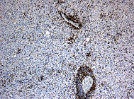
Detection of numerous macrophages (stained brown) in the immunohistochemical staining for CD68 . Chromogenic diaminobenzidine, counterstaining with hematoxylin. Magnification 200x.


therapy
Under suspicion of HSV encephalitis specific therapy with intravenous administration of acyclovir begun and continued for at least three weeks. Foscarnet or ganciclovir are alternatives in the event of a possible resistance of the virus to acyclovir . In order to treat a possible bacterial cause of the symptoms early if the diagnosis has not yet been confirmed, the additional administration of cephalosporins , possibly in combination with ampicillin , makes sense if listeriosis is suspected . A cerebral edema that occurs in the course of the infection is treated with medication; In individual cases, pressure relief was attempted through neurosurgical interventions in order to remove necrosis at the same time.
forecast
If left untreated, HSV encephalitis is fatal in up to 80% of cases. With early use of antivirals, mortality drops to less than 20% of patients. In about half of the surviving children and adults, permanent neurological damage remains despite adequate treatment. Often, memory problems , changes in personality and behavior problems and seizures . A pilot study found evidence of the presence of depressive disorders in ten of 26 patients examined .
literature
- R. Marre, T. Mertens, M. Trautmann, E. Vanek: Clinical Infectiology . Munich / Jena 2000, ISBN 3-437-21740-2 , p. 228 ff.
- N. Suttorp, M. Mielke, W. Kiehl, B. Piece: Infectious diseases . Stuttgart 2004, ISBN 3-13-131691-8 , p. 362 ff.
- RE Levitz: Herpes simplex encephalitis: a review. In: Heart Lung. (1998) 27 (3), pp. 209-212. PMID 9622408
Web links
- Guideline for viral meningoencephalitis of the German Society for Neurology. In: AWMF online (as of October 2005)
Individual evidence
- ↑ E. Schmutzhard: Viral infections of the CNS with special emphasis on herpes simplex infections . In: J. Neurol. (2001) 248 (6), pp. 469-477. (Review) PMID 11499636
- ↑ F. Focher et al.: Sensitivity of monkey B virus (Cercopithecine herpesvirus 1) to antiviral drugs: role of thymidine kinase in antiviral activities of substrate analogs and acyclonucleosides . In: Antimicrob Agents Chemother . (2007) 51 (6), pp. 2028-2034. PMID 17438061
- ↑ Wilson: Viral encephalopathy mimicking functional psychosis. In: Am J Psychiatry. 1976; 133 (2), pp. 165-170. PMID 175665
- ↑ J. Maertzdorf et al.: Herpes simplex virus type 1 (HSV-1) - induced retinitis following herpes simplex encephalitis: indications for brain-to-eye transmission of HSV-1 . Ann. Neurol. (2001) 49 (1), pp. 104-106. PMID 11198277
- ↑ LH Omland et al .: Herpes simplex virus type 2 infections of the central nervous system: A retrospective study of 49 patients . In: Scand. J. Infect. Dis. (2008) 40 (1), pp. 59-62. PMID 17852910 .
- ↑ H. Nakajima et al: Herpes simplex virus type 2 infections presenting as brainstem encephalitis and recurrent myelitis . In: Intern. Med. (1995) 34 (9), pp. 839-842. PMID 8580553
- ↑ F. Frantzidou et al .: Aseptic meningitis and encephalitis because of herpesviruses and enteroviruses in an immunocompetent adult population . In: Eur. J. Neurol. (2008) Jul 12 (Epub). PMID 18637823
- ^ WG Leen et al.: Chronic herpes simplex virus encephalitis in childhood . In: Pediatr. Neurol. (2006) 35 (1), pp. 57-61. PMID 16814088
- ^ S. Love et al.: Chronic granulomatous herpes simplex encephalitis in children . In: J. Neuropathol. Exp. Neurol. (2004) 63 (11), pp. 1173-1181. PMID 15581185 .
- ↑ FD Lakeman, RJ Whitley: Diagnosis of herpes simplex encephalitis: application of polymerase chain reaction to cerebrospinal fluid from brain-biopsied patients and correlation with disease. National Institute of Allergy and Infectious Diseases Collaborative Antiviral Study Group . In: J. Infect. Dis. (1995) 171 (4), pp. 857-863. PMID 7706811
- ^ A. Linde et al .: Specific diagnostic methods for herpesvirus infections of the central nervous system: a consensus review by the European Union Concerted Action on Virus Meningitis and Encephalitis . In: Clin. Diagn. Virol. (1997) 8 (2), pp. 83-104. (Review) PMID 9316731
- ↑ MacCallum et al.: Early diagnosis of herpes simplex encephalitis by brain biopsy. In: Lancet. 1964; 2 (7355), pp. 332-334. PMID 14172330 .
- ^ Whitley et al.: Diseases that mimic herpes simplex encephalitis. Diagnosis, presentation, and outcome. NIAD Collaborative Antiviral Study Group. In: JAMA. 1989; 262 (2), pp. 234-239. PMID 2544743
- ↑ BA Cunha et al: Listeria monocytogenes encephalitis mimicking Herpes Simplex virus encephalitis: the differential diagnostic importance of cerebrospinal fluid lactic acid levels . In: Heart Lung. (2007) 36 (3), pp. 226-231. PMID 17509430
- ↑ G. Feichter, Günter Klöppel, Wolfgang Remmele: Pathology: a textbook and reference book. Springer, Berlin et al. 2012, ISBN 978-3-642-02323-1 .
- ↑ Wiethölter: Inflammatory diseases . In: Pfeiffer et al. (Ed.): Neuropathology. 3. Edition. Springer, Heidelberg a. a. 2002, ISBN 3-540-41333-2 .
- ↑ R. Sánchez-Carpintero et al: Temporal lobectomy in acute complicated herpes simplex encephalitis: technical case report . In: Neurosurgery . (2008) 62 (5), pp. E1174-5. PMID 18580790
- ↑ McGrath et al.: Herpes simplex encephalitis treated with acyclovir: diagnosis and long term outcome. In: J Neurol Neurosurg Psychiatry. 1997; 63 (3), pp. 321-326. PMID 9328248
- ↑ Lahat et al .: Long term neurological outcome of herpes encephalitis. In: Arch Dis Child . 1999; 80 (1), pp. 69-71. PMID 10325763
- ↑ C. Fazekas et al .: Depressive symptoms following herpes simplex encephalitis - an underestimated phenomenon? In: Gen. Hosp. Psychiatry. (2006) 28 (5), pp. 403-407. PMID 16950375