Cat malignant lymphoma
The malignant lymphoma of the cat (syn. Feline lymphosarcoma ) is a malignant (malignant) tumor disease in cats (Felidae), in which mostly solid tumors in the lymphatic organs ( lymphosarcoma form). It is the most common tumor disease in domestic cats , around a third of all neoplasms are malignant lymphomas. Domestic cats are most frequently affected by this specific type of tumor within the species examined so far. The disease is comparable to non-Hodgkin lymphomas in humans. The clinical picture is very variable and depends on the location and size of the tumor. The prospect of a cure with chemotherapy is not always satisfactory, and it is bad for malignant lymphoma of the spinal canal .
etiology
One possible cause of malignant lymphoma is feline leukemia virus (FeLV), a retrovirus . According to a Scottish study, only a third of the affected cats in Europe are FeLV-positive, compared to 70% in North America. However, a typical FeLV antigen , the so-called FOCMA ( feline oncornavirus-associated cell membrane antigen ), can also be detected in most of the seronegative cats . It is assumed that in the majority of cases the organism was able to eliminate the virus, but only after the virus triggered a malignant transformation of the lymphatic cells.
Depending on the presence of FeLV antibody - titer results in a bimodal (bimodal) Age frequency. While most diseases occur in FeLV-positive animals at the age of 3 years, the mean age in FeLV-negative animals is 7 years. In principle, all cat breeds are receptive. However, with the decrease in FeLV as a result of vaccinations, the incidence of malignant lymphomas has not decreased. According to current studies, especially in older animals, only a few animals with malignant lymphoma are also FeLV-positive. More recently, intestinal lymphomas have occurred in particular, and connections with inflammatory bowel disease and cat food and chronic inflammation are currently being discussed.
Another cat-specific virus, the feline immunodeficiency virus (FIV), is also associated with the development of malignant lymphomas to a much lesser extent. A combined effect of both viruses is also being discussed.
Classification
Several classification schemes are possible for malignant lymphoma, which are based on different criteria.
A modified WHO scheme is used after the localization of the tumors . The frequency of the individual forms varies from region to region. The following list is ordered by the number of cases reported worldwide.
- Mediastinal malignant lymphoma : Here the tumor is located in the middle layer ( mediastinum ).
- Gastrointestinal malignant lymphoma : Here are the stomach ( lat. Gaster ) and bowel ( intestine ) site of the primary tumor.
- Peripheral malignant lymphoma : In this form, the peripheral lymph nodes are affected.
- Leukemic leukemia : In this form, there is an increase in white blood cells in the blood ( leukemia ).
- Other malignant lymphomas : This group includes lymphosarcomas that are not located in a lymphatic organ but, for example, in the skin , the central nervous system or the kidneys .
According to the Rappaport classification, a distinction is made between well or poorly differentiated, nodular (nodular) or diffuse as well as large-cell histiocytic and small-cell lymphocytic malignant lymphomas.
After the dignity (about 10%), moderate (35%) and highly malignant (55%) lymphomas distinguished geringgradig.
According to the starting cells, a distinction is made between T-cell tumors, which presumably make up the majority, and B-cell tumors. The latter are mainly found in FIV-associated and gastrointestinal malignant lymphomas. A more precise statement cannot be made here, however, because very few studies have used molecular markers for these lymphocyte subtypes.
clinic
Clinical stages
According to the extent of the disease and the localization-dependent prognosis, a distinction is made between 5 stages, with stage I having the best and stage V the worst.
| Clinical stage | Mark |
|---|---|
| I. | a single tumor of a lymphatic organ |
| II |
multiple lymph nodes in one region |
| III |
Many lymph nodes affected |
| IV | Liver and spleen involvement in addition to a stage I-III feature |
| V | Leukemia or involvement of the bone marrow or central nervous system |
Mediastinal Malignant Lymphoma
A mediastinal malignant lymphoma starts from the lymphatic organs in the middle layer. Often it is the thymus , but the mediastinal ( Lymphonodi mediastinales ) or sternum lymph nodes ( Lymphonodi sternales ) can also be the starting point. This form occurs particularly in the younger, FeLV-positive animals.
Clinically, these lymphomas are generally weak and anorexic . As a result of the constriction of the lungs and the windpipe in shortness of breath or increased breathing rate . As a result of the narrowing of the esophagus , regurgitation or reluctance to drink can occur. The mucous membranes may be pale or bluish in color due to lack of oxygen ( cyanosis ). Changes can occur in the percussion of the rib cage.
An X-ray examination shows shadowing in the precardial (in front of the heart) middle membrane. In terms of differential diagnosis, fluid accumulations ( hydro- , pyo- , chylothorax , feline infectious peritonitis ) and diaphragmatic hernias must be excluded. An ultrasound scan can help you visualize the tumor, especially if it is combined with a build-up of fluid.
A diagnosis can be made using a biopsy of the tumor using fine needle aspiration and a subsequent cytological examination.
Gastrointestinal Malignant Lymphoma
Lymphosarcomas of the gastrointestinal tract or the associated lymph nodes ( Lymphocentrum celiacum , mesentericum craniale and caudale ) are classified in this group. Some authors also classify liver lymphosarcomas in this group. They are rather rare in FeLV-positive animals (10% of cases), but the most common location in older animals and predominantly B-cell tumors. The tumors can appear as solitary nodules, but more rarely also diffuse in the submucosa . About 20% of the cases affect the stomach, the rest the intestines.
The clinical picture depends on the localization. In addition to unwillingness to eat and emaciation, vomiting (especially with solid tumors), diarrhea (diffuse) and, with bleeding , anemia can occur.
Larger solid tumors can possibly already be felt through the abdominal wall. Sonography , endoscopy or laparatomy can provide additional information . The diagnosis is again made by biopsy and cytology.
Peripheral malignant lymphoma
In peripheral malignant lymphoma (also known as multicentric leukosis ), the lymph nodes outside the body cavities are affected. This location is rare in cats, unlike malignant lymphoma in dogs . Clinically, the disease manifests itself unspecifically, mostly painless enlargements of the lymph nodes occur. Later, metastases can occur in the liver, spleen and bone marrow.
The diagnosis is made by biopsy or removal of the entire lymph node and subsequent histological examination.
Leukemic Leukosis
In leukemic leukemia (or lymphoid-leukemic form ), the bone marrow is primarily affected and degenerated lymphocytes circulate in the blood ( leukemia ). Fever, weakness, anorexia, jaundice , fever , anemia, and pale mucous membranes are common.
Other malignant lymphomas

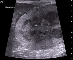
Malignant lymphomas of the kidneys make up around 15% of cases and occur mainly in older animals. The blood values for urea and creatinine are increased. In advanced cases, kidney failure occurs .
Malignant lymphomas of the central nervous system mostly occur in FeLV-positive younger animals or in connection with chemotherapy for renal lymphosarcomas. The tumors mainly occur in the spinal canal and the clinical picture depends very much on the location and extent of compression of the spinal cord . The neurological deficits range from paresis and ataxia to hyperesthesia . Radiographically, in most cases, no changes in the vertebrae can be detected; at most, myelography can provide evidence of a space-occupying process. A number of other central nervous disorders can be excluded from the differential diagnosis, whereby it is best to use the VETAMIN D scheme as a guide.
Malignant lymphomas of the skin are rare and only occur in very old cats (> 11 years). So-called pseudo- abscesses or deeper and more diffusely distributed collections of lymphocytes can occur in the epidermis .
Other possible localizations of malignant lymphomas are the nasal cavity , the conjunctiva and the eye .
therapy
In the case of FeLV-positive animals, treatment is only useful if it is possible to keep them separate from other cats (single indoor cats), as these animals otherwise pose a risk to the cat population.
Chemotherapy has proven to be the drug of choice . In contrast to human medicine, this treatment shows only minor side effects and is well tolerated by the cats. Different protocols for chemotherapy have been proposed so far, but extensive clinical studies on comparability are still pending. A common treatment regimen is the combination of vincristine , cyclophosphamide, and prednisolone . With this so-called COP protocol, remission rates of up to 80% could be achieved; it is particularly promising for mediastinal and peripheral malignant lymphomas. After remission, monthly treatment with doxorubicin or idarubicin is usually carried out to prevent recurrences. Large cell lymphoma intermediate-at gastrointestinal cases proved in 50% and lomustine be effective, well tolerated. The success of chemotherapy cannot be foreseen, however, and the remission rates are lower than in dogs.
Local therapies (surgical removal, radiation ) may be indicated for individual tumors, but chemotherapy to prevent recurrence is also appropriate here.
The treatment of malignant lymphomas of the spinal canal has so far not been very successful.
literature
- KP Richter: Feline gastrointestinal lymphoma. Veterinary Clinics of North America: Small Animal Practice. Volume 33, September 2003, pp. 1083-1098. PMID 14552162 .
- GH Shelton: Feline immunodeficiency virus and feline leukemia virus infections and their relationships to lymphoid malignancies in cats: a retrospective study (1968-1988). In: Journal of Acquired Immune Deficiency Syndromes . Volume 3, 1990, pp. 623-630. PMID 2159993 .
- E. Teske: Hematopoietic Tumors. In: M. Kessler (ed.): Small animal oncology. Parey, Berlin 2000, pp. 523-557. ISBN 3-8263-3236-9 .
Individual evidence
- ↑ a b K. Meichner et al .: Incidence of Feline Leukemia Virus (FeLV) associated malignant lymphoma in the southern German cat population from 1980 to 2008. In: Kleintierpraxis. Volume 56, 2011, p. 35.
- ↑ M. Louwerens et al .: Feline lymphoma in the post-feline leukemia virus era. In: Journal of Veterinary Internal Medicine . Volume 19, May / June 2005, pp. 329-335. PMID 15954547
- ↑ A. Buxbaum et al .: Multicenter malignant lymphoma with conjunctival manifestation in a cat. In: Small Animal Practice. Volume 42, 2000, pp. 699-705.
- ↑ a b S. N. Ettinger: Principles of treatment for feline lymphoma. In: Clinical Techniques in Small Animal Practice. Volume 18, May 2003, pp. 98-102. PMID 12831069 .
- ^ SE Rau, KE Burgess: A retrospective evaluation of lomustine (CeeNU) in 32 treatment naïve cats with intermediate to large cell gastrointestinal lymphoma (2006-2013). In: Veterinary and comparative oncology. Volume 15, number 3, September 2017, pp. 1019-1028, doi: 10.1111 / vco.12243 , PMID 27277825 .
