joint
A joint ( Latin articulatio , ancient Greek ἄρθρον árthron ) from an anatomical point of view is a movable connection between two or more bones .
If these bones fuse, it is called synostosis . In anatomy, a distinction is made between real and fake joints . Of synovial joints is when the joint surfaces are covered with hyaline cartilage, the joint cavity is surrounded by a two-layer joint capsule and a liquid-filled joints between the joint bodies joint space is. A pseudo-joint , on the other hand, refers to the bone area that has remained mobile after an unhealed bone fracture .
Processes and phenomena inside a joint are referred to as intra-articular , e.g. B. intra-articular injection is administration of drugs by puncturing the joint. A joint endoscopy is called an arthroscopy .
False joints (synarthroses)
The fake joints, also known as clinging, include:
-
cartilaginous bone connections ( Juncturae or Articulationes cartilagineae )
- Synchondrosis : connection via hyaline cartilage, for example the costal cartilage , sternum
- Symphysis : Connection via fiber cartilage, e.g. pelvic symphysis , intervertebral discs
-
connective tissue bone connections ( Articulationes fibrosae )
- Suture (suture), for instance between the skull bone
- Syndesmosis (
- Gomphosis (wedging), exclusively for anchoring a tooth in the tooth socket ( articulatio dentoalveolaris )
Real joints (diarthrosis)
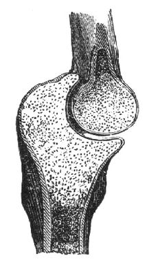
In real joints (also called diarthroses or junctionals synoviales or articulationes synoviales ) there is a gap between the ends of the bones, the joint gap. The joint surfaces are covered by articular cartilage . There is a joint capsule around the joint . It consists of one
- outer membrane fibrosa (tight connective tissue )
- inner synovial membrane (an epithelial-like connective tissue)
The joint or capsular ligaments are strengthened by the fibrous membrane. Joint ligaments can also be independent connective tissue strands, whereby these are located outside the joint capsule (extracapsular ligaments, for example the outer ligament of the knee joint ), in the wall of the joint capsule (for example the inner ligament of the knee joint) or within the joint cavity (intracapsular ligaments, for example the Cruciate ligaments of the knee joint). The latter are covered by a layer of the synovial membrane, which is connected to the joint capsule. Strictly speaking, the cruciate ligaments are therefore intracapsular, but since they cannot be reached directly from the joint cavity without passing through the synovial membrane, this can also be viewed as extra-articular.
The joint capsule completely encloses the joint cavity. It rests loosely on the joint bodies and has appropriate reserve folds ready for joint excursions (very typically observed on the finger joints ). The joint capsule normally only contains a few milliliters of a viscous fluid, the synovia ("joint fluid "), which is a product of the synovial skin of the joint capsule. In the event of inflammation, a joint can bulge with synovia, which is painful and may cause B. can be treated by a puncture. If a joint fills with blood after an accident, it is called hemarthrosis , which must be treated immediately in order to suppress the harmful effects of the blood on the cartilage.
Joint shapes
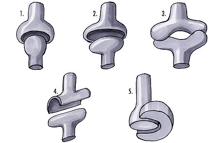
The real joints can be further subdivided according to the shape of the joint surfaces:
- Ball joint (Articulatio sphaeroidea): three-axis, for example shoulder joint, hip joint, metacarpophalangeal joints (with the exception of the thumb); can be moved around the three mutually perpendicular spatial axes: flexion - extension, abduction and adduction, external and internal rotation.
- Ellipsoid or egg joint (Articulatio ellipsoidea): biaxial, for example the first head joint between atlas and skull ; proximal wrist between the radius and the carpal bone; allows both flexion and extension movement as well as side-to-side movement.
- Saddle joint (Articulatio sellaris): biaxial, for example the joint between the carpal bones and the metacarpal bones below the thumb; can be moved around two spatial axes: flexion and extension, abduction and adduction
-
Cylinder joint ( Articulatio cylindrica ), umbrella term for uniaxial joints of the hinge and roll joint type
- Hinge joint (ginglymus): uniaxial, can only be bent ( flexion ) and stretched ( extension ), for example the elbow joint (between humerus and ulna ); the finger joints with the exception of the metatarsophalangeal joints.
- Roll, wheel or pin joint (articulatio trochoidea): uniaxial, for example radioulnar joint (between the radius and ulna) and atlantoaxial joint
- Flat joint or swivel joint (Articulatio plana): no geometric center of movement, also called a flat joint , for example between vertebral processes. Special feature here: two translational degrees of freedom.
- Bicondylar joint or condylar joint (Articulatio bicondylaris): knee joint , biaxial, connection of two condyles with different curvatures: flexion - extension, external and internal rotation (only possible with a flexed knee joint).
- Tight joint ( amphiarthrosis ): for example sacroiliac joint
Joint cracking
The cause of the “cracking” of joints (for example, finger cracking ) has not yet been fully clarified. The most common explanation given is gas bubbles in the synovia , which cause a noise through the formation of bubbles when the pressure is equalized ( cavitation ). Unevenness in the surface of the ankle is also a possible cause.
Habitual finger-cracking is not considered very harmful by experts. It cannot cause arthritis , but it may reduce gripping strength.
Movements in the joints
In order to describe and depict joint excursions, the physical concept of degrees of freedom is also used in anatomy , through which the position of two bodies relative to one another is defined according to a coordinate system in space and which is also translated as "main directions". However, this is misleading because every movement has its antagonistic movement (e.g. internal rotation and external rotation), whereby the directions are opposite, but the plane in which the movements can be represented is exactly one, and this is the degree of freedom . The degree of freedom is perpendicular to the axis of movement.
The range of motion of joints is measured in orthopedics using the neutral-zero method . They are determined from a normal position (“neutral position” or “zero point”) in both directions. The normal position is the posture that the joints assume when standing upright with arms hanging down and feet closed. In this position, for example, the knee is straight and the foot is at right angles to the shin. The range of motion is now measured in both directions, i.e. in the example of the foot the range of dorsiflexion and plantar flexion is determined. The values determined are noted one after the other. Usually the value for the flexion is noted first, then the neutral position comes in the form of a zero and finally the range of motion for the extension is given. In the example of the foot, 30 ° - 0 ° - 40 ° could be logged.
If full mobility is not given due to a contracture and the neutral position is not even reached as a result, the 0 stands as the not reached value of the movement that was not carried out at the beginning or at the end, depending on which value for extension or flexion the passage through Zero prevented. In the case of a hip joint with a flexion contracture of 20 ° (corresponds to a maximum extension of 20 °), the notation could e.g. B. read: 130 ° - 20 ° - 0 °. Since the maximum extension of 20 ° prevented the passage through the neutral point, the zero is behind it, i.e. not within the numbers.
With orthoses , the ranges of motion are also noted using the neutral 0 method. Should z. If, for example, full extension and flexion of the leg due to ligament injuries can be avoided with a knee orthosis, the notation is often 60-10-0. In this setting, the knee can be extended up to a maximum of 10 ° and flexed up to a maximum of 60 °.
Designations for movements
There are names for the different movements of individual body parts, some of which are listed here:
- Abduction = lead away
- Adduction = bring in
- External rotation = turn outwards
- Dorsiflexion
- Extension = stretching
- Flexion = bending
- Inclination
- Internal rotation = turn inwards
- Lateral flexion
- Nutation
- opposition
- Palmar flexion
- Plantar flexion
- Reclination
- Reduction
- Pronation = palm down
- Supination = palm up
- Distal = remote from the body
- Proximal = close to the body
See also
- Arthrolysis
- arthrosis
- Torn ligament
- Ligament stretch
- Joint instability
- Bone inhibition
- Contracture
- dislocation
- sprain
- Arthritis = inflammation of the joints
- Arthrosis = joint wear and tear
- Rickets = softening of the bones due to a vitamin D deficiency
- Osteoporosis = bone loss
- Fracture = break
literature
- TH Schiebler, W. Schmidt (Ed.): Textbook of the entire human anatomy. Cytology, histology, history of development, macroscopic and microscopic anatomy. Taking into account the item catalog . Fourth, expanded and completely revised edition. Berlin / Heidelberg, 1987, ISBN 3-540-16221-6 and ISBN 0-387-16221-6
- F.-V. Salomon: bone connections. In: F.-V. Salomon et al. (Ed.): Anatomy for veterinary medicine. Second edition. Enke-Verlag, Stuttgart 2008, pp. 110-147, ISBN 978-3-8304-1075-1 .
Web links
Individual evidence
- ↑ Johann Schwegler, Runhild Lucius: The human. Anatomy and physiology . Georg Thieme Publishing House. Stuttgart. 2011. p. 175.
- ↑ DL Unger: Does Knuckle Cracking Lead to Arthritis of the Fingers? In: Arthritis and Rheumatism. Volume 41, 1998. pp. 949-950. PMID 9588755 ( Ig Nobel Prize 2009)
- ^ J. Castellanos, D. Axelrod: Effect of habitual knuckle cracking on hand function . In: Annals of the Rheumatic Diseases . 49, 1990, pp. 308-309. doi : 10.1136 / ard.49.5.308 . PMC 1004074 (free full text).
- ↑ Textbook of the entire human anatomy. P. 145.

