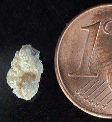
Kidney stone
| Classification according to ICD-10 | |
|---|---|
| N20 | Kidney and ureteral stones |
| N21 | Stone in the lower urinary tract |
| ICD-10 online (WHO version 2019) | |
Kidney stones or nephrolites ( Greek νεφρός nephrós , German ' kidney ' , and λίθος líthos 'stone') are crystalline deposits ( urinary stones ) of the renal pelvis calyx system . When they enter the ureter , they become ureteral stones and can trigger colic . Colloquially, the terms kidney stone and ureteral stone are often used synonymously, although incorrectly. Other names are renal stone or calculus renalis . A collection of many small kidney stones is also called kidney gravel . The medical term for kidney stone disease is nephrolithiasis .
frequency
The incidence of kidney stones in Central and Western Europe is five percent. The ratio of affected men to women is 7 to 5. The disease occurs most frequently between the ages of 30 and 50. In industrialized countries, 20% of men and 7% of women live with an increased risk of stones. If a kidney stone has already occurred, the risk of a relapse (recurrence) is 60%.
Classification
The most common is the division of kidney stones according to their external shape or chemical composition:
-
Classification by shape:
- Valve blocks
- Deer antler stones
- Coral stones
- Pouring stones

- Classification according to chemical composition:
- Calcium oxalate stones (65% frequency)
- Urate -stones (Uric acid stones, 15%)
- Magnesium ammonium phosphate stones ( struvite stones, 11%) occur primarily in connection with infections and are therefore also referred to as infection stones.
- Calcium phosphate stones (9%)
- Cystine stones (approx. 1%)
- Xanthine stones (1%)
- Mixed forms are also possible.
causes
The development of kidney stones depends on many factors which, depending on their severity, lead to concretions of different compositions . Many metabolic processes are still unexplained in this context. At the molecular level, there is an increase in the concentration of poorly soluble ionic compounds or other urine components up to the so-called solubility product being exceeded . As a result, these substances ( salts ) begin to precipitate and form conglomerates which, above a certain size, can no longer pass through the urinary tract .
The increase in the concentration of stone-forming (lithogenic) urine components in the blood and then also in the urine can have many causes. In addition to desiccosis (dehydration) and lack of fluids, diseases that lead to an increased urinary concentration of metabolites or ions , such as hyperparathyroidism , hyperoxalurias , hyperuricemia (increased uric acid , gout ) or certain infectious diseases, are also possible . Abundant dietary intake of purines can increase uric acid levels. There are also kidney problems in which too much calcium phosphate is excreted ( tubular acidosis ). Anatomical peculiarities of the kidney-ureter system such as the horseshoe kidney and ectopic ureter as well as flow obstruction favor the formation of stones.
To an increased formation of kidney stones after a gastric bypass operation, a study with 24 patients suggests where the oxalate - excretion before and after surgery was measured. Before, it was 31 mg per day, then 41 mg. The relative saturation of the urine with calcium oxalate was also significantly increased (1.73 before the bypass operation versus 3.5 after). Every fourth patient developed hyperoxaluria with excretion values of 63 mg per day. Before the operation, no patient was at increased risk of kidney stones.
Antibiotics have been linked to kidney stones. The incidence is particularly high in children.
Symptoms
If stones enter the ureter , they can get stuck in the narrow passages. The resulting spasmodic muscle contractions lead to severe wave-like pain in the affected flank ( renal colic ). As a rule, blood can be seen in the urine or can be detected by laboratory tests. Urine congestion usually occurs and the affected kidney can be damaged. There is a risk of inflammation of the kidneys ( pyelonephritis ) up to uremia or even up to one-sided acute kidney failure ( postrenal kidney failure ). Small stones (maximum diameter up to 6 mm) can come off without any particular discomfort.
Diagnosis
- Physical examination
- Examination of the urine (preferably for traces of blood = hematuria )
- Ultrasound , which can easily miss smaller stones
- X -Kontrastdarstellung both kidneys and the urinary tract (so-called. I v.. - pyelogram ), not suitable for the preparation of urate and Xanthi stones as well as of the rare Indinavir ™ stones
- CT , also shows the so-called non-shadowy concrements that cannot be seen in conventional x-rays
- MRI
- Retrograde contrast agent imaging of the urinary tract
- Endoscopic procedure
- Investigation of asservierten stones in the context of clinical chemistry (so-called stone analysis by infrared spectroscopy )
Ultrasound, urinalysis, and i. v.-pyelogram performed.
therapy
Small kidney stones (less than 6 mm) have a good chance of making their own way through the ureter to the bladder and then through the urethra . Pure urate, struvite and cystine stones can often be dissolved using alkalizing medication ( urolitholysis ). Further measures are:
Percutaneous nephrolitholapaxy (PNL)
This method is mainly used for larger stones. An endoscope is inserted through a small incision in the skin, which is then used to crush the stone using various methods (shock wave, laser, ultrasound). The fragments are then rinsed out. The instruments for this have been miniaturized in recent years.
Ureterorenoscopic Stone Removal (URS)
Such an operative method is used for ureteral stones. A thin tube is inserted through the urethra into the bladder and further into the affected ureter using an optical instrument (similar to a cystoscopy ) . Various devices for breaking up and removing ureteral stones can be inserted through the working channel of the optical instrument. These can be ultrasound, laser, special probes or forceps.
Snare extraction
Because of the high risk of injury, it is only carried out in exceptional cases today. A noose is inserted through the urethra and the doctor tries to pull the stone out. The method is only used when the stone is in the lower third of the ureter. It is no longer mentioned in the EU guidelines for applied medical technology because of the risk of injuring the ureter.
Extracorporeal shock wave lithotripsy (ESWL)
Lithotripsy (from the Greek λίθος 'stone' and τρίβειν 'rub') or ESWL describes the smashing of urinary stones by shock waves generated outside the body . In this process, the focused shock waves are directed onto the stone. In the ideal case, fragments (disintegrates) that can be released spontaneously arise.
The treatment method was successfully carried out for the first time in 1980 by doctors from the Grosshadern University Hospital (Munich, Germany) and engineers and technicians from the Dornier System company ( Friedrichshafen , Germany) (see Dornier kidney stone smashers ). This system is exhibited in the German Medical History Museum in Ingolstadt .
While the first devices (see picture HM 1) still had a tub filled with water in which the patient lay, the newer devices now resemble a modern X-ray device with only one bed. The patient lies on a movable table and is moved to the coupling bellows or the latter to the patient. The coupling bellows consists of a water-filled silicone cover, underneath the acoustic lens and the shock wave generator. This unit is pressed gently against the patient's body in order to establish good contact with the body. In addition, a water-based gel is placed between the surface of the coupling bellows and the skin in order to ensure that the shock waves can pass through without problems. During the treatment, the device automatically detects the position of the stone and corrects the patient's position if the stone moves slightly in the kidney during the shock wave treatment. This ensures that the stone is always in the shock wave center (focal point, focus ) and that the surrounding tissue is spared.
With this procedure, the patient does not need general anesthesia, usually only a mild pain reliever is administered intravenously, the patient remains responsive. The patient is given hearing protection against the noise generated during the treatment (around 3000 low-frequency impulses in 30 minutes). Very often this treatment can also be carried out on an outpatient basis. The burden on the patient is low and, thanks to the targeted bundling of shock waves, less painful than with the first-class devices with a bathtub.
In addition to X-ray cameras, ultrasound devices are also used for stone setting in newer devices. Established methods for generating shock waves are electrohydraulic (spark gap), electromagnetic and piezoelectric generators. Today more than 3000 devices (lithotripter) are used worldwide, around 90% of all kidney stones in industrialized countries are smashed in this way. In 2008 there were around 21,892 ESWL treatments in Germany.
Laser lithotripsy
The destruction of urinary stones has become possible thanks to the development of flexible, thin light fibers with a high damage threshold. Here, an optical quartz fiber is inserted endoscopically under sight until just before the stone to be smashed. If the laser pulse of a dye laser pumped by a flash lamp is now focused on the surface of a kidney stone, the rapid evaporation of the surface material creates a shock wave in the surrounding liquid, which after several shots leads to the stone being shattered. The laser power required for this and the right choice of wavelength at which the absorption of the stone material is maximum depend on the chemical composition of the stone, which can vary. It is therefore useful to know its composition. This can be determined spectroscopically (see: Spectroscopy ) if the fluorescent light emitted by the irradiated stone is collected via a separate fiber and displayed on an optical multi-channel analyzer with low laser energy . A downstream computer can then immediately determine the chemical composition from the spectral distribution of the fluorescence . This was first demonstrated on kidney stones in a water glass ( in vitro ) and then successfully tested on patients ( in vivo ).
Ureteral splint
In almost all of these applications, a catheter (also known as a double J catheter, stent or ureteral splint ) is often left in the ureter to expand and keep open the ureter for a few days or weeks in order to facilitate the natural removal of further stone fragments. The catheter is rolled up for a few centimeters at the upper end in the renal pelvis and at the lower end in the urinary bladder. The double “pigtail” formed in this way fixes the catheter in the ureter. This also protects the ureter, as the stone fragments that leave are partly sharp-edged and the walls of the ureter could be injured.
Roller coaster ride
Using a silicone model with kidney stones of different sizes, US scientists found in 2008 that riding a roller coaster in some cases led to stone loss. The size of the stones played no role in the success rate, but the seat within the row of cars did. The departure rate in the front car was 17 percent and in the last of the five cars 64 percent. The success rate also differed depending on whether it was an upper or lower kidney calculus. The tests could not find out why the stones came off when riding a roller coaster. The trials came after some patients reported passing stones after a roller coaster ride. The tests were carried out on 20 runs without looping, each lasting two and a half minutes. In 2018, the scientists received the Ig Nobel Prize for the experiment .
Metaphylaxis
If a patient has more than one kidney stone, they are more likely to have more stones. To prevent new stones from forming, it is important to find the cause of the stone formation. This is done by means of laboratory, blood and urine tests ordered by a doctor; medical history, occupation and eating behavior must also be included in the tests. The stone should be analyzed for its composition after its removal or its spontaneous loss. To find the cause of the stone formation, the doctor will sometimes examine the urine collected over 24 hours for volume, pH value and the content of calcium , sodium salts , uric acid , oxalate , citrate and creatinine .
- Change in lifestyle
The simplest and most effective way to reduce the risk of new stones forming is to dilute the urine by increasing the daily intake of fluids (mineral water, tea). 2.5 liters of urine should be excreted daily.
Recent studies show that sufficient amounts of calcium in the diet (1000–1200 mg / day) help prevent the formation of oxalate stones. Calcium binds oxalate in the intestine, through which it can be disposed of without any problems. People who are prone to the formation of such stones do not need to limit their consumption of dairy products and other calcium-rich foods. However, it is advised to avoid foods containing calcium-based antacids .
Alkalizing potassium citrate has also proven itself for many decades as protection against kidney stones .
People with acidic urine should avoid meat, fish and poultry, as these foods contain high amounts of purines , the breakdown of which into uric acid lowers the urine pH too much. An increased uric acid level can be a sign of an increased risk of stone formation, which may require medication.
People who have a tendency to form calcium oxalate stones should reduce the following foods rich in oxalate:
- Beets
- chocolate
- coffee
- cola
- nuts
- rhubarb
- spinach
- Strawberries
- Tea (black and to a lesser extent also green)
- Wheat bran
To prevent cystine stones , you need to drink plenty of water, which lowers the cystine concentration in the urine. To do this, you have to drink more than three liters of water every day, a third of it at night.
Herbal medicine
It should be able to dissolve kidney stones with the help of tea made from real bedstraw . A tea infusion of dandelion roots should also help remove the stones. Real cat's whiskers or Orthosiphon relaxes the urinary Harngefäße, works on the inflammation caused by kidney stones, thus reducing overall pain with outgoing stones. A decrease in the nitrogen level in the serum can be observed serologically. Good results are also possible with normal inflammations of the kidneys caused by catarrhed bladder.
research
The presence of the bacterium Oxalobacter formigenes in the intestinal tract can reduce the risk of developing kidney stones by up to 70 percent. This is what a study by a working group at Boston University's Slone Epidemiology Center says . The Boston researchers state that the protective effect of the bacterium is probably based on the metabolism of oxalate in the digestive tract.
literature
- Albrecht Hesse, Dietmar Bach: urinary stones - pathobiochemistry and clinical-chemical diagnostics . (= Clinical chemistry in individual representations . Volume 5). Thieme, Stuttgart 1982, ISBN 3-13-488701-0 .
- Albrecht Hesse, Jörg Joost: Counselor for urinary stone patients . Hippokrates, Stuttgart 1985/1992, ISBN 3-89373-181-4 .
- Albrecht Hesse, Andrea Jahnen, Klaus Klocke: Aftercare for urinary stone patients. A guide for medical practice . Urban & Fischer, 2002, ISBN 3-334-60832-8 .
- Stefan C. Müller, Rainer Hofmann, Kai-Uwe Köhrmann, Albrecht Hesse: Epidemiology, instrumental therapy and metaphylaxis of urinary stones . In: Deutsches Ärzteblatt , 101, issue 19, 2004, pp. A1331–1336.
Web links
- Guideline urolithiasis AK urinary stones of the DGU 2015 [1]
Individual evidence
- ↑ a b Doctors newspaper . July 2, 2008, p. 5, quoted from: JACS 206, 2008, p. 1145.
- ↑ Gregory E. Tasian, Thomas Jemielita, David S. Goldfarb, Lawrence Copelovitch, Jeffrey S. Gerber, Qufei Wu, Michelle R. Denburg: Oral Antibiotic Exposure and Kidney Stone Disease. In: Journal of the American Society of Nephrology. , S. ASN.2017111213, doi : 10.1681 / ASN.2017111213 .
- ↑ S. Müller, R. Hofmann, K. Köhrmann, A. Hesse: Epidemiology, instrumental therapy and metaphylaxis of urinary stones. In: Deutsches Ärzteblatt , 2004, 101 (19), pp. A-1331 / B-1101 / C-1065.
- ↑ Gerold Lingnau : Lithotripter: With shock waves against kidney stones. In: Faz.net. September 29, 2011.
- ↑ Doctors newspaper: Wunderwaffe: roller coaster ride shakes off kidney stones. Retrieved October 8, 2018 .
- ↑ Ig Nobel Prize: Between kidney stones on the roller coaster and sniffing fruit flies . In: ZEIT ONLINE . ( zeit.de [accessed on October 8, 2018]).
- ↑ tagesschau.de: Bottom light: Ig Nobel Prize for kidney stones in the roller coaster. Retrieved October 8, 2018 (German).
- ↑ Pak u. a .: Long-term treatment of calcium nephrolithiasis with potassium citrate. In: J Urol . 1985; 134, pp. 11-19, PMID 3892044 .
- ↑ Volker Fintelmann, Rudolf Fritz Weiss: Textbook of Phytotherapy . Hippokrates-Verlag, Stuttgart 2005, ISBN 3-8304-5345-0 .
- ↑ a b Bacterium Oxalobacter formigenes protects against kidney stones. In: Deutsches Ärzteblatt . March 10, 2008, doi: 10.1681 / ASN.2007101058 .
- ↑ David W. Kaufman et al. a .: Oxalobacter formigenes May Reduce the Risk of Calcium Oxalate Kidney Stones . In: J Am Soc Nephrol . No. 19 , 2008, p. 1197-1203 ( abstract ).


