pineal gland

The pineal gland , epiphysis cerebri or epiphysis for short , anatomically also glandula pinealis ( German name probably after the shape of the cones of the stone pine ( Pinus cembra ); synonymous technical terms see below), is a small, often conical endocrine gland on the back of the midbrain in the Epithalamus (part of the diencephalon ).
In the pineal gland, organ-typical neurosecretory cells , the pinealocytes , produce the hormone melatonin . This neurohormone is formed in the dark and released in the blood and liquor , mostly at night . Melatonin influences the sleep-wake rhythm and other time-dependent rhythms of the body. A malfunction of the pineal secretion can - in addition to a disturbed daily rhythm - cause sexual precocity or delay or inhibition of sexual development.
Synonyms
The pineal gland has several synonymous names:
- Swiss stone pine
- the epiphysis or epiphysis cerebri ( Greek ἐπίφυσις , literally "up-growth", "perched plant", with the Latin addition cerebri - 'of the brain', since the ends of long bones are also referred to as epiphyses )
- the corpus pineale ( Latin , the pine [cone-shaped] body )
- the glandula pinealis (Latin for the pine gland ).
- the pineal organ
- the conarium
anatomy

The pineal gland is an unpaired structure in the middle ( median ) of the brain on the back wall of the III. Ventricle above the quadrilateral plate . It belongs to the circumventricular organs and is anatomically classified as the glandula pinealis among the endocrine glands and assigned to the epithalamus .
In the adult human, the approximately 5-8 mm long and 3-5 mm wide gray-reddish organ has a weight of 80-500 mg, on average about 100 mg. The size of the pineal gland varies in different animal species, also in relation to the size of the entire brain. In some birds it is about one tenth the volume of the brain. Nocturnal animals have smaller pineal glands more often than diurnal animals; in animals that live at high latitudes , such as walruses , the pineal glands are often larger than in animal species in warmer areas of the world, such as elephants . The pineal tissue of mammals is morphologically complex, in some species it does not form a solid pineal body and in others it forms several parts.
Histology and wiring
The pineal gland consists largely of secretory nerve cells ( pinealocytes ) and glial cells .
In the tissue of the pineal gland there are often concentrically layered calcareous concrements of various sizes. These concrements are also known as brain sands ( acervulus , acervuli ) and are visible in the midline of the x-ray of the skull. Their numbers increase with age, and they are found in other parts of the brain as well. So far, brain sand has been detected in many mammals and some birds. The biological meaning is still unclear.
In fish , amphibians , reptiles and many birds, the pineal gland itself is still sensitive to light as the vertex eye ; in mammals , stimuli triggered by light stimuli reach the suprachiasmatic nucleus in the hypothalamus indirectly via the retina and optic nerve . The suprachiasmatic nucleus is the primary chronobiological center of mammals. From here, nerve fibers pass over the dorsal parvicellular subdivision of the paraventricular nucleus , where they accommodate synapses with descending pathways to the spinal cord . These descending pathways lead to the sympathetic root cells ( nucleus intermediolateralis ) in the upper chest marrow. The axons return to the cervical superius ganglion via the neck of the sympathetic nerve (or vagosympathetic trunk ) . From here the information is sent to the epiphysis.
pathology

The pineal cyst is a benign pseudocystic change in the pineal gland that is often found. Tumors of the pineal gland tissue itself - so-called pineal parenchyma tumors, or pinealomas for short - are pineocytoma , a pineal parenchymal tumor of intermediate differentiation and pineoblastoma . In addition, germ cell tumors such as germinoma or a papillary tumor of the pineal region often occur in the pineal gland . Fauchon and colleagues have compiled tumors of the pineal region from various European neurosurgical centers:
| Art | number | percent | Remarks |
|---|---|---|---|
| Germ cell tumors | 96 | 34.4% | Germ cells are usually found in the testes and ovaries . Germ cell tumors can also develop in the pineal region from embryonic remains. |
| parenchymal pineal tumors | 76 | 27.2% | The actual tumors of the pineal gland. |
| Astrocytomas | 52 | 18.6% | Tumors derived from astrocytes , a special type of glial cell . |
| Meningiomas | 20th | 7.2% | Tumors derived from cells in the meninges . |
| Ependymomas | 13 | 4.7% | Tumors that originate from the inner lining of the brain chambers and the neural tube , the ependyma . |
| Oligodendrogliomas | 7th | 2.5% | Tumors derived from oligodendrocytes , a special type of glial cell. |
| Mixed gliomas | 7th | 2.5% | |
| Malignant melanoma | 4th | 1.4% | Melanomas are black skin tumors , but they can also occur inside the body. |
| Metastases | 4th | 1.4% | Once malignant tumors such as lung cancer or breast cancer have settled , they can simulate a pathological enlargement of the pineal gland. |
Pineal tumors can make themselves clinically noticeable through Parinaud's syndrome due to pressure on the quadrilateral plate of the midbrain . Tumors of the pineal region are the most common cause of Nothnagel syndrome .
The pineal gland is easy to see on skull x-rays when it is more calcified - often at an advanced age.
History of the pineal gland and melatonin
Erasistratos of Keos (305-250 BC) and Herophilos of Chalcedon (344-280 BC) were anatomists of the Alexandria school and are (with others of their time) the first anatomists. Erasistratos was interested in the human nervous system, Herophilos was interested in the eye and the human brain. Both believed that the pineal gland was a valve that controlled the flow of our memories.
Galen of Pergamon (130–200), who had also studied in Alexandria and then practiced in Rome , expanded the work of ancient Alexandria with his own anatomical knowledge, but repeatedly referred to the teachings of Hippocrates of Kos . Of Galen's roughly 500 works, 83 have been preserved. He described the location of the pineal gland, its cone-shaped shape and he was already familiar with the frequent calcification of the pineal gland. He was of the opinion that the pineal gland was a kind of valve that would regulate the flow of thoughts in the lateral ventricles ( humoral pathology ). Galen thought the pineal gland was a gland and the pineal region reminded him of the male genital region.
Hindu mystics see the pineal gland as the 7th chakra (crown chakra), which is associated with cosmic energy. It is often assumed that the pineal gland is the 6th chakra, but that this corresponds to the pituitary gland (pituitary gland).
Andreas Vesalius (1514–1564) described the similarity between pineal glands and pine cones .
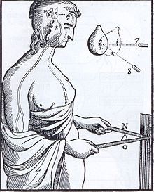
René Descartes (1596–1650), the founder of rationalism , was also interested in the pineal gland. He suspected a direct connection between the eyes and the pineal gland. He saw the main instance of vision in the pineal gland. He believed that this organ coordinates muscle movements with what we see by allowing fluids to flow through tubes between the pineal gland and the muscles (" esprits animaux "). He said of the pineal gland: "There is a small gland in the brain in which the soul functions more specifically than in any other part of the body" ( Les Passions de l'âme , Art. 31).
In 1769, the famous anatomist Morgagni expressed the opinion that calcification of the pineal gland is more common in the mentally ill. Otto Heubner , a pediatrician, observed in 1898 that a boy with early puberty had a pineal tumor . However, it has also been observed that pineal gland tumors can also be associated with delayed onset of puberty. The endocrine function of the pineal gland was also discovered. In 1916 Krabbe considered the production of hormones in the pineal gland. Nils Holmgren , a Swedish anatomist, discovered the similarity between the retina and the pineal gland in frogs and fish in 1918 . The calcifications of the pineal gland visible in the classic X-ray were described by Schüler in 1918.
Kitay and Altschule observed in 1954 that the calcifications of the pineal gland increase with age. At Yale University, the dermatologist Aaron Lerner and his colleague JD Case discovered the structure of melatonin in their search for a drug against vitiligo (white spot disease). In four years of activity they needed around 200,000 bovine pineal glands to isolate melatonin. In the 1960s, Gregory Hill has the pineal gland as a gateway to the inner power in his diskordianistischen religious writing, the Principia Discordia mentioned. Quay discovered the 24-hour rhythm of melatonin secretion in 1964 and in 1965 with his colleagues the synthesis of melatonin in the retina. Russian researchers (Asanova, Rakov?) Described the connection between magnetic fields and melatonin in 1966 . In 1971/72 Konopka and Benzer discovered the per mutation in the fruit fly Drosophila melanogaster . First indications for the functional principle of biochemical or cellular "clocks" (→ chronobiology ). This paved the way for an explanation of the functional principle of cellular clocks. In 1972 Robert Moore and Irving Zucker discovered the location of the "circadian clock" in rats, the nucleus suprachiasmaticus .
In 1973 Piechowak showed the high blood flow to the pineal gland: only the kidney blood flow is higher. In 1978, M. Cohen and co-workers published an article in The Lancet in which they suggested that excessive calcification of the pineal gland could impair its function, which could have implications for the etiology of breast cancer in women. Jenny Redmam showed in 1983 that melatonin injections lead to a shift in their endogenous circadian rhythm in rats and that the timing of the melatonin administration is decisive. The effect of melatonin administration on people suffering from jet lag was investigated by Josephine Arendt in 1986 . The three melatonin receptors Mel1a, Mel1b and Mel1c were 1995 Steve Reppert and DR Weaver cloned . In October 1995, melatonin was classified by the Federal Institute for Consumer Health Protection and Veterinary Medicine (BgVV) as a “medicinally effective substance”, which means that it is no longer freely available as a food supplement in Germany. According to BgVV, melatonin has no nutritional value. Throughout 1995, approximately 50 million melatonin tablets were sold in the United States. The National Institute on Aging (NIA of the NIH) warned in April 1997 against the careless use of melatonin, which is available over the counter in the United States.
Web links
Individual evidence
- ↑ see TA p. 74
- ↑ see TA p. 120
- ↑ CL Ralph: The pineal gland and geographical distribution of animals. In: Int J Biometeorol. 19 (4), 1975, pp. 289-303. on-line
- ↑ L. Vollrath: Comparative Morphology of the Vertebrate Pineal Complex. In: The Pineal Gland of Vertebrates Including Man. Volume 52 of Progress in Brain Research. Elsevier, 2011, doi: 10.1016 / S0079-6123 (08) 62909-X , p. 26.
- ^ PJ Larsen: Tracing autonomic innervation of the rat pineal gland using viral transneuronal tracing. In: Microsc Res Tech . 1999 Aug 15-Sep 1; 46 (4-5), pp. 296-304. PMID 10469465
- ^ A b F Fauchon, A Jouvet, P Paquis, G Saint-Pierre, C Mottolese: Parenchymal pineal tumors: a clinicopathological study of 76 cases . In: International Journal of Radiation Oncology, Biology, Physics . tape 46 , no. 4 , March 1, 2000, ISSN 0360-3016 , p. 959-968 , doi : 10.1016 / s0360-3016 (99) 00389-2 , PMID 10705018 .
- ^ Rudolf Sachsenweger: Neuroophthalmology. 3. Edition. Thieme Verlag, Stuttgart 1983, ISBN 3-13-531003-5 , p. 260.
- ^ Robert A. Zimmerman: Age-Related Incidence of Pineal Calcification Detected by Computed Tomography. (PDF) (No longer available online.) Radiological Society of North America, archived from the original on March 24, 2012 ; Retrieved June 21, 2012 . Info: The archive link was inserted automatically and has not yet been checked. Please check the original and archive link according to the instructions and then remove this notice.
- ↑ Free-running activity rhythms in the rat: entrainment by melatonin . In: Science


