
Development from unicellular to multicellular
Most of the basic lines of development of the eukaryotic domain have produced exclusively unicellular representatives. In the past, when it was not yet clear about their fundamental differences, these were grouped together as “ protists ”. Of the at least about 120 larger clades (cf.) only 36 contain groups that build up multicellular associations (at least in some to many of their species). These are mostly simple cell threads or flat or spherical collections, which are referred to as cell colonies . They usually consist of equally differentiated cells, occasionally two (rarely three) different cell types occur. More highly developed, true multicellular organisms, which are characterized by having numerous different cell types that combine to form complex tissues , exist in six lines of development:
- multicellular animals ( Metazoa )
- Land plants (including mosses) (Embryophyta)
- Red algae of the class Florideophyceae
- Brown algae (" Tange ") of the order Laminariales
- Stand mushrooms (Basidiomycetes)
- Ascomycota (ascomycetes)
The transition to the multicellular way of life was achieved in each of these lines in a unique way independently of the others.
In the remaining 30 or so lines of development with simpler multicellular representatives, multicellular associations have arisen at least 22 times independently of one another, according to the current state of research on the relationships. This suggests that within evolution there is a certain tendency towards multicellular and multicellularity. As before, however, unicellular organisms are not only present today; On the contrary, they have a high density and abundance of species without there being a tendency to be displaced by multicellular / multicellular organisms. While the animals and land plants consist exclusively of multicellular organisms, in all other lines to this day all transitions from single cells to simple colonies and associations to true multicellular cells exist side by side. This would not be possible if the level of organization of the multicellular group were undoubtedly superior in all matters and under all conditions.
Multi-cell: criteria and requirements
The emergence of simple multicellular colonies and aggregates (“clusters”) can easily be achieved through evolution. In fact, it could be artificially generated in the laboratory on a model organism, a unicellular laboratory strain of baker's yeast ( Saccharomyces cerevisiae ) , using suitable selection pressure within a few generations. The development of more complex multicellular life forms with differentiated cells, however, requires the solution of numerous problems.
- All sexually reproducing multicellular cells go back to a single-cell stage, an egg cell, in their development cycle. So apart from secondary mutations , they are genetically identical clones . During cell differentiation, only a few cells are reserved for actual reproduction; the rest perish with the organism they form. So they have to behave altruistically by foregoing their own procreation for the benefit of their relatives.
- The cells of a multicellular organism have to coordinate their behavior, their differentiation and their rate of reproduction so that an organism can arise and function at all. This requires a communication system between the cells.
- For the organism to be able to exist physically at all, the cells have to join together to form cell associations. To do this, they need mechanisms for cell contact and adhesion.
- In addition to cell tissue, all multicellular organisms consist of an extracellular matrix separated from the cells. This must be able to be produced by the cells in a coordinated manner.
In principle, these requirements apply to all multicellular organisms. For more highly developed multicellular cells, further adaptations are also necessary. However, these are initially meaningless for the transition to multicellular life, they only become important when the size and complexity of multicellular organisms increase:
- Differentiated cell associations that work in a division of labor presuppose that some cells have to forego independent food intake. So these have to be fed by others. This requires intercellular transport facilities.
- More highly developed multicellular cells are sealed off from their environment by covering tissue (epithelia) and thus form an internal milieu. This means that they offer the cells inside more uniform, buffered living conditions. However, these cells now have to be transported through other transport devices, e.g. B. be supplied with oxygen.
- More highly developed multicellular cells form numerous different cell lines, although the genetic material is the same in all of their cells. So you need a complex control of development and differentiation.
Advantages and disadvantages of simple multicellular cells
Multi-celled organisms have always evolved from unicellular organisms. The resulting, first multicellular organisms must therefore initially have been very simply structured, quite small organisms, which initially could not have had too many different cell types. In order for this stage to arise and be selected by evolution, it must have had a selective advantage over its immediate unicellular relatives. For this consideration, the advantages of large, highly differentiated multicellular cells are completely irrelevant. Since the historical transition to multicellular cells took place a long time ago (in the case of all complex multicellular organisms already in the Precambrian ), small, poorly differentiated multicellular cells living today are particularly important for comparison with their unicellular relatives.
The following benefits have been documented for simple multicellular organisms: They are against a number of predators ( predators better protected), for example, unicellular predators or those with be filtered diet. The green alga Scenedesmus acutus lives z. B. in the laboratory mostly as a single cell, in the field as a cell colony. It could be shown that the presence of daphnia ("water fleas") as predators in the same water body triggers the transition to colony formation. You can add nutrients such as B. store phosphate more effectively for times of emergency and shortage (e.g. Volvox ).
The advantages are offset by some significant disadvantages of multicellular organisms. This applies in particular to the simple and first, as yet poorly differentiated, multicellular cells. By clarifying the relationship, z. In the case of the relatives of the green alga Volvox , for example , it can indeed be shown that more simply structured or even single-cell lines have emerged from multicellular lines. First of all, the cells of a newly emerging multicellular cell have the same problem that arises on a different organizational level for social animal species. When some cell lines give up direct reproduction in favor of germ cells, they behave altruistically , that is, they accept disadvantages for themselves that benefit other cells. Altruistic systems are always threatened by “fraudsters” who take advantage of the benefits without contributing anything themselves. In more highly developed cell associations, the individual cells quasi give up their individuality in favor of the newly emerging organism. Independent reproduction occurs here only pathologically (as cancer ); this occurs in various forms in all multicellular organisms from almost all evolutionary lines (so it is not limited to vertebrates with humans). In the case of more simply structured multicellular cells, whose cells might also be viable on their own or which would be able to produce viable offspring instead of the germ cells, the altruistic task of self-reproduction assumes corresponding advantages for other (usually closely related) carriers of the same genetic material. Another disadvantage of a growing organism is that the simple distribution of substances by diffusion can no longer be sufficient to supply all cells, which is why special, possibly complex transport devices have to be newly developed.
So how was it possible that multicellular organisms could arise when they initially had only a few selection advantages over unicellulars and their higher development depended on numerous evolutionary innovations, without which they could not realize these advantages? It seems to be a classic “ chicken and egg problem ”. To clarify this question, living, simple and hardly differentiated multicellular cells or their relatives that have remained unicellular are examined as model organisms . Classic model organisms for the evolution of multicellular life forms, such as B. the green alga Volvox , the cellular "slime mold" Dictyostelium discoideum or the colony-forming frilled flagellate Monosiga brevicollis are presented in more detail below. In short, the solution to the riddle can be summarized as follows: Numerous adaptations, of which it was traditionally assumed that they were only present and useful in multicellular animals and therefore had to be newly developed by them, were actually already present in single-celled organisms, this becomes pre-adaptation or also Called exaptation . As a result, the hurdle to the development of the multicellular cells was lower than long thought.
Death and cell death
The main difference between unicellular and multicellular organisms is that in multicellular organisms only certain cells, the germ cells, are involved in reproduction. The remaining cells (called somatic cells , from the Greek σῶμα soma "body") perish with the organism. In addition to the death of the individual who leaves a corpse , and partly as the cause for this, the individual cells of a multicellular cell are also not able to live and divide indefinitely. Their life often ends actively with a kind of built-in "suicide program" with programmed cell death , usually in the form of apoptosis . Many cells fulfill their role in the organism even only when they are dead, such as B. the water conducting tissue ( xylem ) of land plants or the horny cells of the epidermis, hair and horns of vertebrates. Single-cell organisms, on the other hand, are considered "potentially immortal". Their life does not end through aging and death, but rather through cell division in (usually) two daughter organisms, otherwise only through enemies or accidents.
Recent research has now shown that unicellular organisms can also die, i.e. that they are not necessarily potentially immortal, as the simplistic view suggested. This even applies to prokaryotes . The enzymes used, the caspases (in plants: metacaspases) are homologous to those of the multicellular organisms in many unicellular organisms, and the other mechanisms, as far as is already known, have homologous formation in multicellular organisms. At least in some cases, the mechanism appears to have been acquired through horizontal gene transfer , making it difficult to interpret the actual timing. But today it is highly probable that the basic mechanism is older than multicellular life forms. The suicide program is triggered by signals from conspecifics, so it is a form of coordinated behavior in groups or colonies of single cells.
Various hypotheses have been put forward about the biological meaning of programmed cell death in single cells. It often occurs during oxidative stress or under nutrient deficiency conditions. It may be used to provide conspecifics (usually close relatives due to the clonal growth) with nutrients when the conditions for their own reproduction are no longer sufficient. However, this connection is speculative and in individual cases no convincing advantage has yet been found. As with multicellular cells, the defense against pathogens and infections probably also plays a role.
adhesion
In order for a potential multicellular cell not to simply fall apart, bridges or bonds between its cells are necessary (see under cell contact ). This process is called adhesion .
Adhesion mechanisms are fundamentally different between organisms whose cells form a cell wall and those with "naked" cells. Multicellular cells with cell walls usually “stick” their cell walls to one another with polysaccharides such as pectins or hemicelluloses . Direct contact between the cells is created through gaps in the wall (so-called pits ) via connections called plasmodesmata . In multicellular red algae, so-called “pit connections” are formed according to similar principles, but in a completely different way. Similar pores are found between fungal cells.
Animal cells make cell contacts mainly via membrane proteins. These either span the cell membrane and are then in direct contact with elements of the cytoskeleton , or they are anchored in the membrane via so-called GPI anchors (with the substance glycosylphosphatidylinositol). The adhesion molecules of animals can be divided into five families, the cadherins , integrins , selectins , proteoglycans and immunoglobulin -like ones. By fully deciphering the genome of the single cell Monosiga brevicollis , a member of the Choanomonada , which has been recognized as the closest relative of multicellular animals, we know that most of them are common to multicellular animals and their unicellular relatives. This can only be explained by the fact that they were created in evolution before the first multicellular animals developed. Monosiga has 23 cadherins, 17 integrins (all with alpha subunits), 5 immunoglobulins and 12 so-called C-type lectins. So there is not just one adhesion molecule, but a whole battery of them. The exact biological role of these molecules in Monosiga is still unclear. However, it seems highly probable that it has something to do with cell contacts in him too.
The sponges are among the most simply structured multicellular animals, whose cells have striking similarities to the unicellular choanomonads . From experiments, some of which were carried out as early as the 19th century, it is known that the cell aggregates of sponges can regenerate spontaneously if the cells have been separated beforehand - for example by streaking the organism through a sieve. Complex proteoglycans have been identified as the main adhesion molecules that bring about this organization.
Signals
The communication of cells in multicellular organisms (see under signal transduction ) is a prerequisite for almost every form of multicellular life. In multicellular cells, adhesion and signal exchange are often linked and are often mediated by the same molecules.
The simplest multicellers get by with little signal exchange. In the case of the green alga Volvox rousseletii, for example , no direct signal exchange is necessary for swimming to the light, which requires the coordinated work of the flagella of all cells of the spherical cell cluster. Here each cell reacts independently to the incidence of light. The different size and light sensitivity of the cells at the front and rear end result in easy coordination of movement. But even the cell colonies from Volvox , which in addition to the sex cells only consist of a single cell type, cannot do without communication. They have developed special cyclins (of type D) that coordinate cell reproduction and reproduction.
In multicellular animals, an essential class of signaling molecules is the tyrosine kinases . Their development was therefore long considered a key feature of multicellular animals. From the decoding of the genome of the choanomonads Monosiga brevicollis it has now become clear that this not only has tyrosine kinases but also a greater variety of families of these enzymes than all Metazoa . Most of them have no direct orthologs in multicellulars, so they were developed convergently (on the same basis) . Not much is known about the special role of the tyrosine kinases in Monosiga ; previous evidence suggests signal exchange. Cell cultures of the species only achieved a much lower density when the kinases were inhibited .
Extracellular matrix
All multicellular organisms do not consist exclusively of cells. A large part of the organism is formed by substances that are secreted outwards by cells in a highly coordinated manner. This extracellular matrix is of great importance in both simply built, poorly differentiated multicellular cells and in complex, highly differentiated organisms. They give form and shape to simple, poorly differentiated multicellular cells. The spherical cell colony of the green alga Volvox consists of 99 percent extracellular matrix. Simply structured multicellular animals also get their structure from them. The jelly-like umbrella or the polyp of a cnidarians or a comb jellyfish consists essentially of cell-free substance ( mesogloea ), through which thin layers ( epithelia ) of active cells are kept in shape. The body of a sponge is also mainly formed by a cell-free matrix ( called mesohyl here ) (it is what remains of the organism in a real bath sponge when it is used). In addition, cells called scleroblasts secrete calcareous or pebbly skeletal needles ( spicules ) in most sponges .
Such deposits are also widespread in unicellular organisms. They include enzymes for digesting food (e.g. invertases , proteases , siderophores ), toxins and antibiotics , but also structural molecules that give them e.g. B. help to form so-called biofilms on surfaces of all kinds. This can be selectively maintained in that e.g. B. all those involved benefit from increased aggregations etc. and that neighbors among microorganisms are very likely to be close relatives due to the clonal growth (cf. under relative selection ). In the case of iron bacteria that produce siderophores, however, it has been shown that, in the long term, it is not uncommon for “fraudsters” who do not contribute anything to prevail in the population at the expense of their altruistic relatives. Under unfavorable conditions, however, the growth of the entire colony suffers compared to the cooperative type.
Cell types
The simplest multicellular organisms consist of cells that are completely identical to one another. Simple cell differentiations, however, already occur in prokaryotes . The cyanobacterium ("blue-green algae") Nostoc forms simple cell threads from interconnected cells. Scattered into the thread are thick-walled cells with a different morphology , called heterocysts . Their task is to fix nitrogen from molecular nitrogen from the air, the key enzyme of which, nitrogenase, is sensitive to oxygen. Heterocysts have to be supplied with assimilates by the photosynthetically active cells and supply these in turn with nitrogen compounds. Differentiated multicellularity is also found in spore-forming myxobacteria , e.g. B. the much studied Myxococcus xanthus observed. However, it is generally rare in prokaryotes. It is certainly not by chance that the multicellular representatives have significantly larger genomes than their unicellular relatives. The protein-coding genome of Myxococcus xanthus is one of the largest known of all prokaryotes and even surpasses some simple eukaryotes.
Multicellular animals have more different cell types. The lowest number reported is four (possibly five) for the simplest multicellular Trichoplax adhaerens . Most metazoa have between 10 and 25 different cell types. Much higher numbers are only given for vertebrates. For the model organism Homo sapiens , 411 are currently named, of which 145 different types of neurons (nerve cells) alone . The comparison of the different cell types is possible between animals today by "molecular fingerprint" of the different tribes belong; it used to be unclear whether similar cell types can be traced back to common ancestors or whether they had become more similar to one another in a secondary manner through convergent evolution. The general trend that emerged here is that multiply specialized cells in simple multicellulars must have developed into increasingly specialized ones in complex multicellulars. For example, cnidarians have cells that belong to the outer covering tissue (epithelium), which both absorb sensory stimuli from the environment and are capable of contracting (contracting) like muscle cells. These functions are divided between different cell types in higher multicellular cells (in cells homologous to them).
The differentiation has not progressed because the first multicellular cells gradually acquired new functions and new cell types (and tissues or organs) acquired for them. Rather, the original cells already possessed most of the later functions, combined in a single cell. These “all-rounders” then differentiated themselves based on the division of labor. The few cell types of the simply structured multicellular organisms - and even more so the unicellular ones - are therefore not simpler than those of the more complex multicellular organisms, on the contrary, they are more complex.
Model organisms
The genus Volvox

Structure of Chlamydomonas
1. Flagella
2. Vacuole
3. Cell wall
4. Eye spot
5. Cell nucleus
6. Cup-shaped chloroplast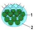
Structure of the green alga Gonium
1. chlamydomonas-
like cell
2. Jelly
Construction of the Volvox
1. chlamydomonas-
like cell
2. daughter sphere
3. plasma bridges
4. jelly
5. reproductive
cell 6. body cell


The Volvocales order is often used as a model for researching the first stages of multicellularity, because it contains the complete series from real unicellular cells, few cell colonies to real multicellular cells, the cells of which also have easily comparable shape and morphology. The Volvocales are green algae ( Chlorophyta ), whose line of development separated more than a billion years ago from that which led to the multicellular land plants ( Embryophyta ). It is assumed that the (completely independent) transition to multicellularity took place here later (probably in the Triassic ).
The species Chlamydomonas reinhardtii is by far the best researched representative of the unicellular Volvocales . It is a soil-living alga that is believed to only occur in eastern North America, but is now cultivated in laboratories around the world. Related species are common all over the globe. Chlamydomonas species living in the ground are extremely tough against adverse environmental conditions and can hardly be killed. They live in moistened mud or small collections of water on the soil surface. If the conditions are favorable, the ( haploid ) cells divide asexually. Here, two mitotic divisions are carried out in quick succession within the cell wall of the mother cell; the four daughter cells then separate and continue to live as single cells.
If the conditions are unfavorable, the cells transform into gametes (of the same size, therefore not males and females, but called plus and minus forms). After mating, they form extremely resistant permanent spores (so-called zygospores), which wait for better conditions in dried out algae crusts. They even occur in desert soils. Due to the strongly changing environmental conditions in the soil, the cells must be able to tolerate a wide variety of environmental conditions. This means they can continue to live under oxygen-free ( anaerobic ) conditions. If they don't get any light, switch to a heterotrophic diet (based on acetic acid ). Missing nutrients such as phosphorus and nitrogen can be enriched with special enzymes. Their resistance to heavy metals is so high that they are being considered for the biotechnological purification of wastewater contaminated with heavy metals. Chlamydomonas cells are motile by two flagella ; they swim towards the light ( phototaxis ), the direction of which they can see through an eye spot.
Since Chlamydomonas cells divide several times in quick succession after having increased significantly in size, the transition to the cell colony stage seems very obvious. For this it is only necessary that the daughter cells remain attached to one another after division. Within the Volvocales there are a number of genera that resemble such Chlamydomonas colonies. The number of cells is (depending on the division steps) 4, 8, 16 or a multiple thereof. Disc-shaped cell colonies from such cells are z. B. placed in the genera Gonium and Eudorina .
From a certain size of the cell colony, this simple organization is no longer sufficient. All larger representatives form hollow spherical shapes. These are placed in the genus Volvox . In fact, this transition took place several times independently of one another. The species of the “genus” are therefore not closely related to one another, but rather belong to different lines of development that have convergedly developed the same shape. In all cases, the problems encountered with the increase in size were resolved in a similar manner.
Volvox organisms consist of two types of cells. In addition to the Chlamydomonas -like somatic cells, germ cells have appeared here, which are used exclusively for (asexual) reproduction (sexual reproduction continues to exist with the formation of permanent spores). The increase in size could be directly responsible for this differentiation. Because of the cell organization (the centrioles , which organize the spindle apparatus during cell division, also serve as the starting point for the flagella), the cells can no longer swim long after they have started to divide. If too many cells divide simultaneously within a Volvox ball, it sinks to the bottom and perishes (the balls are heavier than water and have to swim continuously). With some Volvox species such as B. Volvox carteri , the generative cells are also larger due to asymmetrical division and can deliver larger embryos, which have a starting advantage. At Volvox, the embryos are not released to the outside but to the inside of the hollow sphere. This is because they would not be viable at first as their flagella point inward. You must first turn yourself inside out in a process called inversion. The finished embryos contain all the cells of the adult sphere. They are initially still connected to each other by cytoplasmic bridges which (in most Volvox species) are later pinched off.
The formation of the multicellular organism Volvox therefore requires a rather complex sequence of steps despite its simplicity. Cell differentiation has to be controlled, the extracellular matrix (which has developed from the cell wall of Chlamydomonas ), which takes up 99 percent of the spherical volume, has to be deposited in a coordinated manner, the embryo has to go through the complicated inversion ... This requires considerable advantages for this development who have built up a corresponding selection pressure . What these advantages are is not entirely clear. The storage of nutrients (especially phosphorus) in the extracellular matrix as storage for times of shortage and the better protection that the size gives against many predators (e.g. filter feeders ) are mentioned most frequently .
While Chlamydomonas is widespread almost everywhere, Volvox is only found in relatively few habitats, especially ponds and very nutrient-rich (layered) lakes, where it can become more common than Chlamydomonas . In these mostly murky waters, it may also benefit from its greater mobility through numerous coordinated whipping flagella. Volvox has its advantages over Chlamydomonas especially in a habitat that offers particularly favorable development opportunities. The advantages are by no means unambiguous, especially under unfavorable environmental conditions: Due to the molecular pedigree of the Volvocales, it is now clear that there are numerous reversions in the evolution of the group, i.e. H. Reverse developments to less cellular forms must have come. If the division of labor of the cells offers too few advantages, especially with smaller cell colonies, the somatic cells (which otherwise traditionally forego their own reproduction) can go back to independent reproduction.
Social amoeba
The Eumycetozoa are a group of organisms that have long been referred to as fungi (" slime molds "), but with which they actually have nothing in common except a superficial similarity of the fruiting bodies. Today their membership of the Amoebozoa has been proven without a doubt. The group of Dictyostelia with the model organism Dictyostelium discoideum has been researched best . Dictyostelia include around 100 species. They occur worldwide in soils of all kinds, but have a distribution focus in forest soils. The species Dictyostelium discoideum is restricted to the Chinese coastal region, Japan and eastern North America. Molecular studies have shown that the three conventional genera of Dictyostelia, which are differentiated according to the shape of the fruiting bodies, are not natural relatives; the (overall monophyletic ) group can be divided into four lines of development, each comprising members of several genera. However, a revision has not been made, above all in order not to have to change the introduced species names.
Dictyostelia feed as unicellular, swarm-forming amoebas on soil bacteria, which they devour by phagocytosis . In this phase of life they reproduce asexually through simple cell division. The bacteria are recognized by their metabolic products, especially folic acid . The amoeba are mobile by the eversion of pseudopodia , which are formed (among other things) by a cell skeleton made of actin and myosin fibers. These loose swarms of protozoa can develop a multicellular fruiting body when their food starts to run out. First, the amoeba combine to form an aggregated, mobile structure that is called “slug” or pseudoplasmodium and crawls around for a while. If a favorable place is reached, it changes into a fruiting body called a sporocarp . The fruiting body consists of a multicellular stem which, depending on the species, has one or more spherical spore containers on top. Although a distinction has been made between up to five cell types, it basically consists of two different cell lines. The spore cells ultimately form new amoeba; the stem cells perish with the fruiting body. The formation of the multicellular fruiting body from up to around 100,000 unicellular, independent amoebas requires intensive communication between the individual cells, which has been extensively researched in Dictyostelium . In addition, altruistic cells are also involved here: the amoebas that form the stem cells sacrifice their own ability to reproduce in favor of their relatives. In Dictyostelium, behavior coordination and differentiation are based on chemical communication. Each cell emits signaling substances that are perceived by other species and trigger oriented behavior ( chemotaxis ) and cell differentiation.
In Dictyostelium discoideum and related species (but not in other Dictyostelia) the communication between the cells is largely based on G-protein receptors with the signaling molecules cAMP and cGMP . It corresponds to the communication between many cells of multicellular animals, e.g. B. the leukocytes of the blood of mammals. The amoebas continuously measure both the bacterial concentration in their environment (presumably via the folic acid content) and the cell density of the amoeba surrounding them (via a glycoprotein). When there is a lack of food, a few pacemaker cells begin to release cAMP. This causes the amoeba to join together to form a dense aggregate by moving forward following the substance gradient to the location of the highest concentration. The cell mass exudes an enveloping cellulose-containing mucus layer on the outside. The cell mass finally elongates and begins to crawl around as a “slug”, influencing not only chemical but also optical and temperature stimuli. The cells of the front end of the "slug" ultimately differentiate into the stalk and die. The other cells (about four fifths of the total number) form a spherical spore body, which serves as a stage of spread and duration. Cell differentiation is controlled by additional signal molecules called DIF (differentiation-inducing factor).
Although Dictyostelium cells often form extensive aggregates of clonal single cells in the feeding phase, mixed (“chimeric”) fruiting bodies are occasionally formed. However, the cells are able to recognize relatives with whom they prefer to associate. Still, like all altruistic collectives, they have to deal with “cheaters”; In this case, these are cell lines that only form spores and do not contribute to the formation of the stalk. Such fraudulent strains can be interspersed in natural populations. At least some tribes have developed the ability to recognize and exclude them.
Multicellular animals
The multicellular animals or Metazoa form (according to molecular pedigree analyzes) a monophyletic community of descent, that is, they all descend from a common ancestral form. Research tries to clarify these so-called "primeval metazoa" primarily by comparing the construction plans and developing the simplest metazoa that are still alive today. The most important candidates for the most pristine still living Metazoa are the sponges ( Phylum Porifera).
Since the discovery of the unicellular or colony-forming collared flagellates (choanoflagellates), their extraordinary resemblance to the collared flagellate cells ( choanocytes ) of the sponges has been noticed . Both consist of a cell body with a single flagella at one end. This is surrounded by a "collar" made of cell processes ( microvilli ). Nutrition takes place when food particles are swirled by the flagella to the collar, where they stick and are phagocytosed . As early as the 19th century it was therefore assumed that sponges, and thus all Metazoa, could have developed from colonies of flagellates. This model is still the most important hypothesis about the origin of the Metazoa. The more mobile “higher” metazoa such as the bilateria could then have developed through a stage corresponding to the larval stage of the sponges, the planula , while the fixed, filtering stage was abandoned ( neoteny ).
Closer examination of the choanoflagellates, for example the species Salpingoeca rosetta , already revealed a previously unexpected diversity of cell types and life forms. Salpingoeca occurs in five different stages (some of which were previously understood as different species or even genera): a) Fixed (sessile) single cells that sit in a cup-shaped, stalked shell or "theca", b) slowly swimming single cells, c) fast swimming single cells without “collar”, therefore very similar to sperm , d) spherical (“rosette-shaped”) and e) chain-shaped, freely swimming cell colonies. Five differently differentiated cell types can be distinguished. These are no less than in many sponges. The main difference here is that they do not occur simultaneously in an organized multi-cell, but one after the other. The transition between the forms is triggered by environmental stimuli, for example a molecule that occurs in bacteria species used as food triggers the formation of rosette-shaped colonies.
Various models have been proposed for the transition from a hypothetical ancestral form that resembles a colony of today's choanomonads to a multicellular cell:
- "Gastraea": According to this, a simple, hollow spherical cell colony (or "Blastaea") would have formed initially. In a second step it turned inside out, creating a two-layer spherical colony. This would then have differentiated. This hypothesis dates back to the 19th century; it goes back to the biologist Ernst Haeckel , who developed it in analogy to the blastula and gastrula stages of embryogenesis . A modern version comes from z. B. by Claus Nielsen
- “Placula”: According to this hypothesis, the multicellular cells would have emerged from a soil-living colony, the cells of which would have been differentiated into a lower, mobile and an upper part serving for food intake. These would later have differentiated into two cell layers. This hypothesis, first formulated by the zoologist Otto Bütschli , gains credibility through the discovery of the Placozoa , which resemble this stage.
- “Synzoospore”: According to this hypothesis, the first mobile stages (which would possibly resemble the Blastaea) would not have been free living organisms, but only a mobile larval stage of a differentiated, fixed (sessile) cell colony or an aggregate of unicellular cells. This would have arisen because, at first, single-cell swarmers (zoospores) remained attached to one another as a bandage. The theory was put forward by the Russian biologist Alexej Alexejewitsch Zachvatkin .
See also
Individual evidence
- ↑ Sina M. Adl, Alastair GB Simpson, Mark A. Farmer, Robert A. Andersen, O. Roger Anderson, John A. Barta, Samual S. Bowser, Guy Bragerolle, Robert A. Fensome, Suzanne Fredericq, Timothy Y. James , Sergei Karpov, Paul Kugrens, John Krug, Christopher E. Lane, Louise A. Lewis, Jean Lodge, Denis H. Lynn, David G. Mann, Richard M. McCourt, Leonel Mendoza, Øjvind Moestrup, Sharon E. Mozley-Standridge , Thomas A. Nerad, Carol A. Shearer, Alexey V. Smirnov, Frederick W. Spiegel, Max FJR Taylor (2005): The New Higher Level Classification of Eukaryotes with Emphasis on the Taxonomy of Protists. Journal of Eukaryotic Microbiology 52 (5): 399-451. doi : 10.1111 / j.1550-7408.2005.00053.x
- ↑ : SM Adl, BS Leander, AG Simpson, JM Archibald, OR Anderson, D. Bass, SS Bowser, G. Brugerolle, MA Farmer, S. Karpov, M. Kolisko, CE Lane, DJ Lodge, DG Mann, R. Meisterfeld, L. Mendoza, O. Moestrup, SE Mozley-Standridge, AV Smirnov, F. Spiegel (2007): Diversity, Nomenclature, and Taxonomy of Protists. Systematic Biology 56 (4): 684-689. doi : 10.1080 / 10635150701494127
- ↑ Adl, SM, Simpson, AGB, Lane, CE, Lukeš, J., Bass, D., Bowser, SS, Brown, MW, Burki, F., Dunthorn, M., Hampl, V., Heiss, A. , Hoppenrath, M., Lara, E., le Gall, L., Lynn, DH, McManus, H., Mitchell, EAD, Mozley-Stanridge, SE, Parfrey, LW, Pawlowski, J., Rueckert, S., Shadwick, L., Schoch, CL, Smirnov, A. and Spiegel, FW (2012), The Revised Classification of Eukaryotes. Journal of Eukaryotic Microbiology, 59: 429-514. doi : 10.1111 / j.1550-7408.2012.00644.x
- ^ A b Andrew H. Knoll (2011): The Multiple Origins of Complex Multicellularity. Annual Review of Earth and Planetary Sciences 39: 217-239. doi : 10.1146 / annurev.earth.031208.100209
- ↑ Sean B. Carroll (2001): Chance and necessity: the evolution of morphological complexity and diversity. Nature 409: 1102-1109.
- ^ William C. Ratcliff, R. Ford Denison, Mark Borrello, Michael Travisano (2012): Experimental evolution of multicellularity. Proceedings of the National Academy of Sciences USA Vol. 109 No.5: 1595-1600. doi : 10.1073 / pnas.1115323109
- ^ Lewis Wolpert & Eörs Szathmáry (2002): Evolution and the egg. Nature 420: 745.
- ^ A b Dale Kaiser (2001): Building a multicellular organism. Annual Revue of Genetics 35: 103-123.
- ↑ > Richard K. Grosberg & Richard R. Strathmann (2007): The Evolution of Multicellularity: A Minor Major Transition? Annual Review of Ecology, Evolution, and Systematics 38: 621-654. doi : 10.1146 / annurev.ecolsys.36.102403.114735
- ↑ David L. Kirk (2005): A twelve-step program for evolving multicellularity and a division of labor. BioEssays 27: 299-310.
- ↑ Richard E. Michod (2007): Evolution of individuality during the transition from unicellular to multicellular life. Proceedings of the National Academy of Sciences USA vol. 104, suppl. 1: 8613-8618. doi : 10.1073 / pnas.0701489104
- ↑ C. Athena Aktipis, Amy M. Boddy, Gunther Jansen, Urszula Hibner, Michael E. Hochberg, Carlo C. Maley, Gerald S. Wilkinson (2015): Cancer across the tree of life: cooperation and cheating in multicellularity. Philosophical Transactions of the Royal Society B 370: 20140219. doi : 10.1098 / rstb.2014.0219
- ^ Raymond E. Goldstein (2009): Evolution of Biological Complexity. Seminaire Poincare XII: 75-88.
- ↑ a b J.C. Ameisen (2002): On the origin, evolution, and nature of programmed cell death: a timeline of four billion years. Cell Death and Differentiation 9: 367-393.
- ↑ a b Marıa Segovia, Liti Haramaty, John A. Berges, Paul G. Falkowski (2003): Cell Death in the Unicellular Chlorophyte Dunaliella tertiolecta. A Hypothesis on the Evolution of Apoptosis in Higher Plants and Metazoans. Plant Physiology Vol. 132: 99-105.
- ^ A b Daniel J. Franklin, Corina PD Brussaard, John A. Berges (2006): What is the role and nature of programmed cell death in phytoplankton ecology? European Journal of Phycology 41 (1): 1-14. doi : 10.1080 / 09670260500505433
- ↑ a b Hanna Engelberg-Kulka, Shahar Amitai, Ilana Kolodkin-Gal, Ronen Hazan (2006): Bacterial Programmed Cell Death and Multicellular Behavior in Bacteria. PLoS Genetics Volume 2, Issue 10, e135. doi : 10.1371 / journal.pgen.0020135
- ^ Kris Vleminckx (2001): Adhesive Specificity and the Evolution of Multicellularity. Encyclopedia of Life Sciences doi : 10.1038 / npg.els.0001273
- ↑ Monica Medina, Allen G. Collins, John W. Taylor, James W. Valentine, Jere H. Lipps, Linda Amaral-Zettler, Mitchell L. Sogin (2003): Phylogeny of Opisthokonta and the evolution of multicellularity and complexity in Fungi and Metazoa. International Journal of Astrobiology 2 (3): 203-211. doi : 10.1017 / S1473550403001551
- ↑ Nicole King, M. Jody Westbrook, Susan L. Young, Alan Kuo, Monika Abedin, Jarrod Chapman, Stephen Fairclough, Uffe Hellsten, Yoh Isogai, Ivica Letunic, Michael Marr, David Pincus, Nicholas Putnam, Antonis Rokas, Kevin J. Wright, Richard Zuzow, William Dirks, Matthew Good, David Goodstein, Derek Lemons, Wanqing Li, Jessica B. Lyons, Andrea Morris, Scott Nichols, Daniel J. Richter, Asaf Salamov, JGI Sequencing, Peer Bork, Wendell A. Lim, Gerard Manning, W. Todd Miller, William McGinnis, Harris Shapiro, Robert Tjian, Igor V. Grigoriev, Daniel Rokhsar (2008): The genome of the choanoflagellate Monosiga brevicollis and the origin of metazoans. Nature 451: 783-788 doi : 10.1038 / nature06617
- ↑ Gradimir N. Misevic, Camille Ripoll, Jonathan Norris, Vic Norris, Yann Guerardel, Emmanuel Maes, Gerard Strecker, Pascal Ballet, Yannis Karamanos, Lazar T. Sumanovski, Octavian Popescu, Nikola Misevic (2007): Evolution of multicellularity in Porifera via self assembly of glyconectin carbohydrates. in: MR: Custódio, G. Lôbo-Hajdu, E. Hajdu, G. Muricy (editors): Porifera research: biodiversity, innovation and sustainability. Série Livros 28, Museu Nacional, Rio de Janeiro. PDF ( Memento of the original from October 29, 2013 in the Internet Archive ) Info: The archive link was inserted automatically and has not yet been checked. Please check the original and archive link according to the instructions and then remove this notice.
- ↑ Noriko Ueki, Shigeru Matsunaga, Isao Inouye, Armin Hallmann (2010): How 5000 independent rowers coordinate their strokes in order to row into the sunlight: Phototaxis in the multicellular green alga Volvox. BMC Biology 8: 103 download
- ↑ a b Simon E. Prochnik, James Umen, Aurora M. Nedelcu, Armin Hallmann, Stephen M. Miller, Ichiro Nishii, Patrick Ferris, Alan Kuo, Therese Mitros, Lillian K. Fritz-Laylin, Uffe Hellsten, Jarrod Chapman, Oleg Simakov, Stefan A. Rensing, Astrid Terry, Jasmyn Pangilinan, Vladimir Kapitonov, Jerzy Jurka, Asaf Salamov, Harris Shapiro, Jeremy Schmutz, Jane Grimwood, Erika Lindquist, Susan Lucas, Igor V. Grigoriev, Rüdiger Schmitt, David Kirk, Daniel S. Rokhsar (2010): Genomic Analysis of Organizational Complexity in the Multicellular Green Alga Volvox carteri. Science. 329 (5988): 223-226. doi : 10.1126 / science.1188800
- ↑ Gerard Manning, Susan L. Young, W. Todd Miller, Yufeng Zhai The protist, Monosiga brevicollis, has a tyrosine kinase signaling network more elaborate and diverse than found in any known metazoan. Proceedings of the National Academy of Sciences USA 105 (28): 9674-9679.
- ↑ Annalisa M. VanHook (2008): Facilitating Multicellularity. Science Signaling Vol. 1, Issue 29: p. ec262 doi : 10.1126 / scisignal.129ec262
- ↑ Nicole King, Christopher T. Hittinger, Sean B. Carroll (2003): Evolution of Key Cell Signaling and Adhesion Protein Families Predates Animal Origins. Science 301: 361-363.
- ↑ Review in Stuart A. West, Stephen P. Diggle, Angus Buckling, Andy Gardner, Ashleigh S. Griffin (2007): The Social Lives of Microbes. Annual Review of Ecology, Evolution, and Systematics 38: 53-77. doi : 10.1146 / annurev.ecolsys.38.091206.095740
- ↑ Jan Mazek & Anne O. Summers (2008): General Characteristics of Procaryotic Genomes. In: Ying Xu & J Peter Gogarten (editors): Computational Methods for Understanding Bacterial and Archaeal Genomes. Series on Advances in Bioinformatics and Computational Biology: Volume 7. ISBN 978-1-86094-982-1
- ↑ a b Detlev Arendt (2008): The evolution of cell types in animals: emerging principles from molecular studies. Nature Reviews Genetics 9: 868-882.
- ↑ Bernd Schierwater, Danielle de Jong, Rob DeSalle (2009): Placozoa and the evolution of Metazoa and intrasomatic cell differentiation. International Journal of Biochemistry & Cell Biology Volume 41, Issue 2: 370-379. doi : 10.1016 / j.biocel.2008.09.023
- ↑ Graham Bell & Anne O. Moors (1997): Size and complexity among multicellular organisms. Biological Journal of the Linnean Society 60: 345-363.
- ↑ MK Vickaryous & BK: Hall (2006): Human cell type diversity, evolution, development, and classification with special reference to cells derived from the neural crest. Biological Reviews 81: 425-455. doi : 10.1017 / S1464793106007068
- ^ Daniel W. McShea (2002): A complexity drain on cells in the evolution of multicellularity. Evolution 56 (3): 441-452.
- ^ Matthew D. Herron, Jeremiah D. Hackett, Frank O. Aylward, Richard E. Michod (2009): Triassic origin and early radiation of multicellular volvocine algae. Proccedings of the National Academy of Sciences USA Vol. 106 No.9: 3254-3258. doi : 10.1073 / pnas.0811205106
- ↑ a b Elisabeth H. Harris (2001): Chlamydomonas as model organism. Annual Review of Plant Physiology and Plant Molecular Biology 52: 363-406.
- ↑ Cristian A. Solari, Aurora M. Nedelcu, Richard E. Michod (2003): Fitness, Life-history, and the Evolution of Complexity in Volvocalean Green Algae. PDF
- ↑ David L. Kirk (2005): A twelve-step program for evolving multicellularity and a division of labor. BioEssays 27.3: 299-310.
- ↑ Cristian A. Solari, Richard E. Michod, Raymond E. Goldstein (2008): Volvox barberi, the fastest swimmer of the Volvocales (Chlorophyceae). Journal of Phycology 44: 1395-1398. doi : 10.1111 / j.1529-8817.2008.00603.x
- ^ Joel L. Sachs (2008): Resolving the first steps to multicellularity. Trends in Ecology and Evolution Vol.23 No.5: 245-248. doi : 10.1016 / j.tree.2008.02.003
- ↑ Steven L. Stephenson & Alan Feest (2012): Ecology of Soil Eumycetozoans. Acta Protozoologica 51 (3): 201-208. doi : 10.4467 / 16890027AP.12.016.0762
- ↑ AR Swanson, EM Vadell, JC Cavender (1999): Global distribution of forest soil dictyostelids. Journal of Biogeography 26: 133-148. doi : 10.1046 / j.1365-2699.1999.00250.x
- Jump up ↑ Pauline Schaap, Thomas Winckler, Michaela Nelson, Elisa Alvarez-Curto, Barrie Elgie, Hiromitsu Hagiwara, James Cavender, Alicia Milano-Curto, Daniel E. Rozen, Theodor Dingermann, Rupert Mutzel, Sandra L. Baldauf (2006): Molecular Phylogeny and Evolution of Morphology in the Social Amoebas. Science Vol. 314 no. 5799: 661-663. doi : 10.1126 / science.1130670
- ↑ Carole A. Parent & Peter N. Devreotes A Cell's Sense of Direction. Science Vol. 284 no. 5415: 765-770. doi : 10.1126 / science.284.5415.765
- ↑ a b Si. I. Li & Michael D. Purugganan (2011): The cooperative amoeba: Dictyostelium as a model for social evolution. Trends in Genetics Vol. 27, No. 2: 48-54. doi : 10.1016 / j.tig.2010.11.003
- ↑ Werner EG Müller (2001): How was metazoan threshold crossed? The hypothetical Urmetazoa. Comparative Biochemistry and Physiology Part A 129: 433-460.
- ↑ a b Claus Nielsen (2008): Six major steps in animal evolution: are we derived sponge larvae? Evolution & Development 10 (2): 241-257.
- ↑ Mark J. Dayel, Rosanna A. Alegado, Stephen R. Fairclough, Tera C. Levin, Scott A. Nichols, Kent McDonald, Nicole King (2011): Cell differentiation and morphogenesis in the colony-forming choanoflagellate Salpingoeca rosetta. Developmental Biology 357 (1): 73-82. doi : 10.1016 / j.ydbio.2011.06.003
- ↑ Rosanna A Alegado, Laura W Brown, Shugeng Cao, Renee K Dermenjian, Richard Zuzow, Stephen R Fairclough, Jon Clardy, Nicole King (2012): A bacterial sulfonolipid triggers multicellular development in the closest living relatives of animals. eLife 2012; 1: e00013 doi : 10.7554 / eLife.00013
- ↑ but is not identical with it. see. M. Carr, BSC Leadbeater, R. Hassan, M. Nelson, SL Baldauf (2008): Molecular phylogeny of choanoflagellates, the sister group to Metazoa. Proceedings of the National Academy of Sciences vol. 105 no. 43: 16641–16646 doi : 10.1073 / pnas.0801667105
- ↑ Bernd Schierwater, Michael Eitel, Wolfgang Jakob, Hans-Jürgen Osigus, Heike Hadrys, Stephen L. Dellaporta, Sergios-Orestis Kolokotronis, Rob DeSalle (2009): Concatenated Analysis Sheds Light on Early Metazoan Evolution and Fuels a Modern '' Urmetazoon ' 'Hypothesis. PLoS Biology 7 (1): e1000020. doi : 10.1371 / journal.pbio.1000020
- ↑ Kirill V. Mikhailov, Anastasiya V. Konstantinova, Mikhail A. Nikitin, Peter V. Troshin, Leonid Yu. Rusin, Vassily A. Lyubetsky, Yuri V. Panchin, Alexander P. Mylnikov, Leonid L. Moroz, Sudhir Kumar, Vladimir V. Aleoshin (2009): The origin of Metazoa: a transition from temporal to spatial cell differentiation. BioEssays 31: 758-768. doi : 10.1002 / bies.200800214