T lymphocyte

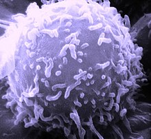
T lymphocytes, or T cells for short, form a group of white blood cells that are used for immune defense . Together with the B lymphocytes, T lymphocytes represent the acquired (adaptive) immune response . The T in the name stands for the thymus in which the cells mature.
Like all blood cells, T cells are made in the bone marrow . From there they migrate into the thymus, where MHC receptors are formed on their surface. Through an initial positive selection, followed by a negative selection , all those who react to the body's own proteins or who cannot recognize the body's own MHC receptors are rejected. The remaining, remaining T cells can then only recognize exogenous antigens and thus do not fight the body itself. The proteins in the selected cell membranes, also called T cell receptors (TCR), can then - similar to those of B lymphocytes antibodies produced - recognize exogenous substances. In contrast to antibodies, however, T cells only recognize foreign substances if their antigens are bound to their MHC on the surface of other cells. Free antigens are only recognized by T lymphocytes if they are actively shown by so - called antigen - presenting cells (so-called MHC restriction).
function
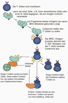
T cells migrate through the organism and constantly monitor the membrane composition of the body cells for pathological changes. Foreign or changed substances on the cell surface can be caused, for example, by a virus infection or by a mutation of the genetic material. If one of the presented MHC-I or MHC-II molecules on the surface of the diseased cell exactly matches the individual receptor of a passing T-cell like a key in the associated lock, and if at the same time a costimulant (e.g. the surface protein B7 ) is presented, the T cell goes into the activated state by activating certain genes of the cell nucleus . Antigen receptor and co-receptor together form the activation signal. The cell grows and differentiates into effector and memory cells . Depending on the type of cell, the effector cells have different functions. Killer T cells (characterized by the CD8 receptor ) destroy the diseased cell directly; T helper cells (with CD4 receptor ) sound the alarm with soluble messenger substances ( cytokines ) and attract additional immune cells. Regulatory T cells prevent excessive attacks on intact body cells, so they help with self-tolerance . T cells are thus responsible for cell-mediated cytotoxicity , for controlling the humoral immune response and, last but not least, for many allergic reactions . The strength of the various reactions depends on the stimulating antigen, the type of presenting cell, and other factors, some of which are still unknown.
In the thymus, a lymphatic organ , the new T cells are prepared for their various functions. A classification can be made on the basis of the surface antigens CD4 and CD8: CD4 + T lymphocytes are regarded as helper cells; their receptor recognizes MHC class II molecules. CD8 + T lymphocytes are considered cytotoxic T cells; their receptor recognizes antigens that are presented by almost all body cells via MHC class I molecules. In fact, only the most common combination of T cell phenotype and function is shown; there are also CD4 + cytotoxic T cells and CD8 + T helper cells. CD4 + T cells are mainly found in the peripheral blood and in lymphatic tissues with a high blood supply, such as the parafollicular regions of the lymph nodes , spleen and tonsils . In contrast, CD8 + T cells are more likely to be found in the bone marrow and in the lymphatic tissues of the gastrointestinal mucosa , the respiratory organs and the urinary tract .
Naive (inactive) T cells are constantly moving between the blood and these lymphoid tissues. For this purpose, they have little amoeboid mobility and are equipped with cell adhesion molecules and receptors for chemokines . They leave the blood stream by means of diapedesis through the walls of the postcapillary venules. From there they migrate through the tissue and return to the blood with the lymph via the thoracic duct , which opens into the left vein corner. Another possibility is that the lymphocytes migrate through the walls of a high endothelial venole (HEV) into a secondary lymphatic organ.
Special functions of the T lymphocytes
Immune cells play an important role in controlling bone metabolism. They can release substances that promote the breakdown of the bone matrix by osteoclasts . Under estrogen deficiency , T lymphocytes were stimulated to produce TNF-α and RANKL in the mouse model ; this could have contributed to the development of bone mineral loss in the test animals. Thymusless mice from which the ovaries had been removed did not suffer any bone loss despite the lack of hormones. In athymic nude mice and rats, the bone turnover rate is generally lower. Osteoprotegerin from activated T lymphocytes stimulates the osteoclastic breakdown of the bone substance and could be involved in the development of bone and joint diseases.
Structure and differentiation of related cell types
T and B lymphocytes are spherical cells similar in size to red blood cells ; their diameter in humans is about 7.5 µm. They cannot be differentiated from one another microscopically or electron microscopically. Only by means of immunohistochemistry can marker proteins such as CD3 , which is characteristic of T lymphocytes, and CD19 , which is specific for B lymphocytes , be represented. The chromatin in the round or slightly indented, non-lobed cell nucleus is dense, lumpy and vividly colored. The plasma border around the core is narrow and can hardly be seen with a light microscope . The numerous lysosomes can be seen as azurophilic granules. The cell substance contains plenty of free ribosomes . The Golgi apparatus is smaller than that of the reticular cells.
The T-cell antigen receptor (TCR)
Each TCR on peripheral T cells is bound to a CD3 receptor molecule. The CD3 receptor directs the activation signal into the cell interior. It binds to both TCRαβ and TCRγδ. The extent of the reaction of the receptor complexes with the antigen-MHC complexes depends on the concentration of both partners; H. their density on the cell membranes involved, and the specific affinity of the TCR. With the crystallography , the three-dimensional structure of the TCR was clarified. The antigen-binding, hypervariable V-region resembles the corresponding V-domain of antibodies. These molecule segments in the form of exposed loops determine the antigen specificity of the receptor and are also called complementarity determining regions CDR.
Like the antigen receptor of the B lymphocytes, the TCR belongs to the immunoglobulin gene superfamily. Two of four possible protein chains (denoted by α, β, γ, δ) are linked by disulfide bridges . Usually the TCR is an αβ heterodimer, more rarely a γδ heterodimer. So two subpopulations of T cells can be distinguished. The α chains weigh 43–49 kilodaltons , the β chains 38–44 kilodaltons , the γ chains 55–60 and the δ chains approx. 40 kilodaltons. At 15 nm, the complex of receptor and MHC is small compared to other membrane proteins. The genes for the α and δ chains are nested at the same location on chromosome 14q11-12, the γ chain gene is on chromosome 7p15 and the β chain gene is on chromosome 7q32-35. The arrangement of the genes does not make it possible for a cell to simultaneously develop receptors as γδ and αβ heterodimers.
In the blood circulation and in the lymphatic organs, 95–98% of the T cells belong to the αβ subpopulation. CD4 + and CD8 + T cells belong to it. γδ-T cells are predominantly (up to 50%) to be found in epithelial tissues such as the skin, the intestinal mucosa or the genital organs , i.e. on the body surfaces.
Lower forms
While in the 80s T cells were divided into the two forms T helper ( CD4 +) and T suppressor cells ( CD8 +), we now know the high "plasticity" of T cells, which are divided into others Change subtypes or develop characteristics of several subtypes, depending on the soluble mediators present, and which can produce cytokines and interleukins specific for the subtype. Subtypes are e.g. B. T1, T2, T9 or T17.
T helper cells
T cells with a helper function secrete different cytokines and can be classified according to whether these messenger substances are involved in the cell-mediated immune response or whether the humoral immune response of the B lymphocytes is stimulated. The presence of IL-12 and interferon -γ (IFN-γ) induces differentiation to the TH1 cell, while IL-4 and IL-6 promote differentiation to the TH2 cell. For example, CD4 + lymphocytes belong to the first group (type 1) which secrete interferon-γ (IFN-γ), IL-2 , and TNF-α . CD4 + lymphocytes, which produce the cytokines IL-4 , IL-5 , IL-6 , IL-10 and IL-13 , are classified as type 2. The same distinction can also be made for secreting CD8 + T cells and for those with a γδ T cell antigen receptor. There are also T-helper cells with a mixed cytokine pattern called Type0 T cells.
The differences between type 1 T cells and type 2 T cells were first described by Tim Mosmann in 1986 .
Cytotoxic T cells
The cytotoxic T cells (CTL, obsolete killer cells ) are usually characterized by CD8 + -αβ heterodimers on the surface. They recognize antigens presented on MHC-I molecules, especially virally infected cells and tumor cells. CTL trigger programmed cell death in the defective cells via their physiological signaling pathways (Fas / FasL ; Perforin / Granzyme ).
Regulatory T cells (T Reg )
The intensity of the immune response must be constantly monitored, on the one hand to destroy the cancer cells and pathogens , but to suppress autoimmunity against normal tissue. In addition, the post-production and maturation of the leukocytes must be kept constant. Part of the control mechanisms are exercised by regulatory T cells (obsolete suppressor T cells ): via cytokines such as IL-10 and TGF-β , through the capture of antigens, growth and differentiation factors, through CTLA4-mediated limitation of clonal expansion by B cells, and by killing excess T cells via Fas / FasL-mediated signals. The regulatory T cells are further subdivided on the basis of their cytokine profiles, for example into (CD4 + -CD25 + T-reg cells, T R 1 cells, T H 3 lymphocytes and NKT cells, CD8 + regulatory cells).
T memory cells
Memory T cells form a kind of “immunological memory” by remaining in the blood after they have been activated. If the same pathogen is infected again, the original activation is restored. The presence of memory cells increases the multiplication of antigen-specific T cells by 10 to 100 times. The memory role can be exercised by both CD4 + and CD8 + T memory cells.
NK T cells
The natural killer T cells are a small number of cytotoxic αβ T cells that have no antigen-specific receptors, but can still recognize the presentation of MHC-I antigen complexes. They are T cells, so you shouldn't confuse them with the natural killer cells of the unspecific immune system. NK-T cells are characterized by the molecule NKR-P1A , a lectin- like protein, on their surface. Further markers expressed by NK cells are CD56 , Neural cell adhesion molecule-1 (NCAM-1) and CD57 . These cells also produce the cytotoxic effector molecules perforin and granzym. The task of the NK-T cells is to be in the control of autoimmune diseases.
γδ antigen receptor positive T lymphocytes
γδ T cells make up only a small percentage of the T cells in blood and lymphoid organs, but are very prominent in the skin and many epithelial tissues. Its essential feature is a T cell receptor, which consists of the γ and δ subunits. The γδ TCR is significantly less varied than the αβ TCR; only a few of the many theoretically possible combinations of the various gene segments are used. The binding partners of the γδ TCR are still largely unknown, but it is probably mainly the body's own molecules (the αβ TCR usually recognizes the antigens of pathogens).
In terms of development and function, γδ T cells differ significantly from αβ T cells. They leave the thymus in a pre-activated state, which enables a quick reaction and rapid release of active substances. Activation can probably also take place independently of the γδ TCR by cytokines. γδ T cells recognize tissue damage and changes (such as cancer) and then activate both innate and acquired components of the immune system.
T-lymphocyte-bound diseases
Congenital immune defects
Inherited immunodeficiencies affecting both T cells and B cells, i.e. H. Damage to the cellular and humoral immune responses are called severe combined immunodeficiency (SCID). The affected children must be cared for in an environment with as few germs as possible and only have a chance of survival in the long term after a successful bone marrow transplant .
The DiGeorge syndrome prevents the development of epithelial tissue in the thymus of the fetus. As a result, the T cells cannot mature and the cellular immune response is greatly reduced.
Patients with naked lymphocyte syndrome develop leukocytes and thymus cells without MHC-II molecules and thus a lack of CD4 + T lymphocytes.
Acquired immune deficiencies
Acquired immunodeficiencies can be caused by various diseases, malnutrition, harmful effects of the environment or therapeutic measures.
Infections
The human immunodeficiency virus (HIV) infects CD4 + T lymphocytes, dendritic cells and macrophages, which leads to the immunodeficiency disease AIDS . The viruses HTLV I and HTLV II can attack T-lymphocytes in humans and primates and cause various diseases, including adult T-cell leukemia and tropical spastic paraparesis .
Allergic reaction
A hypersensitivity reaction , or specific allergy , is when an inappropriate immune reaction is triggered against the body's own tissue or against an actually harmless antigen (dust, pollen , food or pharmaceuticals). Among the four types according to Coombs and Gell, T cells are mainly involved in type I (immediate type) and type IV (delayed type). In the immediate type, an excessive T 2 response is marked, in the case of the delayed allergy a persistently excessive activity of the T 1 cells and thus persistent inflammation.
Autoimmune diseases
Autoimmune diseases are chronic diseases caused by immune reactions against the body's own antigens. In type I diabetes mellitus, for example, it has been observed that insulin- specific CD8 + T cells attack β cells in the pancreas . Even with the rheumatoid arthritis autoreactive T cells have been demonstrated. According to the widespread theory of multiple sclerosis , this disease is also initiated by activated T cells, which destroy the myelin sheaths of nerve cells .
Drug effects
Certain drugs can cause desirable and undesirable immune deficiencies. After organ transplants , there is a risk of transplant rejection, which involves both cellular and humoral immune reactions. The focus is on the T-cell reaction against allogeneic and xenogeneic MHC molecules in foreign tissue. Studies show three mechanisms: acute rejection by CD8 + T cells, chronic rejection by CD4 + T cells, and damage to the blood vessels supplying the transplant. All three mechanisms can be permanently suppressed by immunosuppressive drugs. Growth-inhibiting, cell-killing drugs such as cytostatics and radiation with ionizing radiation can also damage white blood cells and especially T-lymphocytes.
Oncological disease patterns
Degenerate T cells are the starting point of a group of tumor diseases ( malignant lymphomas ) and acute lymphoblastic leukemia , which often affects patients in childhood.
The occurrence of T lymphocytes in other living things
In invertebrates (such as protozoa , sponges , annelids and arthropods ) there are neither lymphocytes nor lymph nodes. In vertebrates , lymph nodes only appear in birds and mammals , whereas lymphocytes are more in the family tree in cartilage and bony fish as well as amphibians and reptiles .
Research history
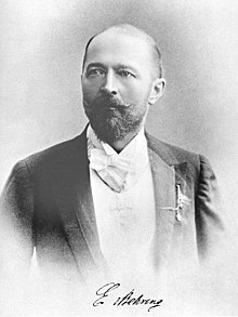
At the beginning of the 20th century, the subject of scientific debate was whether the immunity of some people against infection is based on cellular or humoral processes. The zoologist Elias Metschnikow (1845–1916) observed that mobile cells accumulated around a thorn pricked into a starfish . Metschnikow assumed that these cells eat invading bacteria ( phagocytosis theory ). In contrast, scholars such as Emil Adolf von Behring (1854–1917) took the view that immunity is produced by substances dissolved in blood serum . In 1888, Bering found that the multiplication of anthrax bacteria was prevented by serum from resistant rats, but not by serum from guinea pigs susceptible to anthrax. Only the serum of those guinea pigs that had previously been infected with this germ was effective against Vibrio metschnikovii , and their serum, in turn, was not effective against other germs. With this, Behring was also able to refute Hans Buchner , who had believed that the blood serum had an unspecific bactericidal activity. Together with Kitasato , Behring developed his theory of humoral immunity and what is known as "blood serum therapy".
Belgian researchers (Denys, Lecleff and Marchand ), but especially Almroth Wright and SR Douglas, were able to resolve the apparent contradiction between the two theories around 1903. Wright and Douglas found phagocytosis-promoting substances in serum, which they called opsonins - today's antibodies - which linked cellular and humoral processes. According to the instruction theory published by Linus Pauling in 1940 , antigens formed an instruction, according to which blood cells transform a universal immunoprotein into a suitable specific antibody.
Deviating from this, Niels Jerne ( clone selection theory ) and Paul Ehrlich ( side chain theory ) took the view that all immunoglobulins are already preformed and that the correct one is selected by the introduced antigen. Frank MacFarlane Burnet then recognized that it was not the circulating antibodies that were selected, but rather individual immunocompetent cells, which then multiply to form a specifically producing clone ( Nobel Prize 1960). During embryonic life, somatic mutations give rise to countless variants of possible antigen receptors; at the same time, those cells that carry receptors for the body's own antigens are eliminated again.
By 1926, the role of lymphocytes in the rejection of exogenous tissue was recognized. Gowans described in 1964 that such lymphocytes are available everywhere, as they switch from the breast duct into the blood and then back into the tissue via the secondary lymph organs. The special importance of the thymus was discovered in leukemic mice in 1968. In the mid-1960s, a distinction was made between B and T lymphocytes. Jerne described their interaction in antibody production in 1974 (Nobel Prize 1984). In 1975, Kisielow and co-workers differentiated cytotoxic from non-cytotoxic T cells. In 1976, Rolf Zinkernagel and Peter Doherty showed that the T cell is only activated when the triggering antigen is presented at the MHC. In 1982 it was possible to synthesize a mAb that recognized T-cell lymphoma cells in mice. The surface structures and TCRs of the T cells have been described in more detail using T cell hybridomas and leukemic T cell lines. In 1979, Kung found the CD3 proteins on the side of the T cell antigen receptor; their biochemical characterization followed in 1984 by a research group from Cox Terhorst.
The T-cell receptor was described in mice in 1983 as a 45-50 kDa heterodimer with an α and a β chain. In the following year, the mRNA isolation of the human TCR was also successful, for the first time with the help of the cloning of β chains of the human and mouse TCRs. A few years later a second TCR, similar to the αβ TCR, was found - the γδ T cell antigen receptor. The MHC restriction of the T cell antigen receptor was also described for the first time in 1986. TCR genes can be involved in chromosomal mutations that activate cancer-promoting oncogenes . With the help of molecular samples from TCR, genes could be identified that play a role in the development of leukemia and lymphoma. 1988-89 it was shown that CD8 is the receptive partner for those antigens that are presented at the MHC-I. The memory of CD4 and CD8 cells has been described.
literature
- GA Holländer: Immunology, basics for clinics and practice . 1st edition. Elsevier, Munich 2006, ISBN 3-437-21301-6 .
- MJ Owen, JR Lamb: Immune Recognition . Thieme, Stuttgart 1991, ISBN 3-13-754101-8 .
- I. Jahn: history of biology, theories, methods, institutions, short bibliographies . 3. Edition. Gustav Fischer, Jena 1998, ISBN 3-437-35010-2 .
- A. Wollmar, T. Dingermann: Immunology, Basics and Active Ingredients . With the collaboration of I. Zündorf. Wissenschaftliche Verlagsgesellschaft, Stuttgart 2005, ISBN 3-8047-2189-3 .
- O. Bucher, H. Wartenberg: Cytology, histology and microscopic human anatomy . 11th edition. Hans Huber, Bern / Stuttgart / Toronto 1992, ISBN 3-456-81803-3 .
- K. Munk: Basic studies in biology and zoology . Gustav Fischer, Heidelberg / Berlin 2002, ISBN 3-8274-0908-X .
Web links
- uni-tuebingen. de / List / List01. aspx? subject = Immunobiology Lecture Immunobiology of T lymphocytes Video recordings of a lecture. From TIMMS, Tübingen Internet Multimedia Server of the University of Tübingen .
Individual evidence
- ↑ YY Kong, H. Yoshida, I. Sarosi, HL Tan, E. Timms, C. Capparelli, S. Morony, AJ Oliveira-dos-Santos, G. Van, A. Itie, W. Khoo, A. Wakeham, CR Dunstan, DL Lacey, TW Mak, WJ Boyle, JM Penninger: OPGL is a key regulator of osteoclastogenesis, lymphocyte development and lymph-node organogenesis . In: Nature . tape 397 , no. 6717 , January 28, 1999, p. 315-323 , PMID 9950424 .
- ^ S. Cenci, MN Weitzmann, C. Roggia, N. Namba, D. Novack, J. Woodring, R. Pacifici: Estrogen deficiency induces bone loss by enhancing T-cell production of TNF-alpha . In: Journal of Clinical Investigation . tape 106 , no. 10 , November 2000, pp. 1229-1237 , PMID 11086024 .
- ↑ M. Girardi: Immunosurveillance and immunoregulation by γδ T cells . In: J. of Investigative Dermatology . No. 126 , 2006, pp. 25-31 , PMID 16417214 .
- ↑ Stefanie Sarantopoulos: Allogenic stem-cell transplantation - a T-cell balancing ACT. In: New England Journal of Medicine . Volume 378, Issue 5, February 1, 2018, pp. 480-482; doi: 10.1056 / NEJMcibr1713238
- ↑ Tim Mosmann, H. Cherwinski, MW Bond, MA Giedlin, RL Coffman: Two types of murine helper T cell clone. I. Definition according to profiles of lymphokine activities and secreted proteins . In: Journal of Immunology . tape 136 , no. 7 , 1986, pp. 2348-2357 ( jimmunol. Org / cgi / content / abstract / 136/7/2348 abstract ).
- ^ M. Bonneville, RL O'Brien, WK Born: Gammadelta T cell effector functions: a blend of innate programming and acquired plasticity. . In: Nat. Rev. Immunol. . tape 10 , 2010, p. 467-478 , PMID 20539306 .
- ↑ AC Hayday: Gamma delta T cells and the lymphoid stress surveillance-response. . In: Immunity . tape 31 , 2009, p. 184-196 , PMID 19699170 .
- ↑ György Nagy, Joanna M. Clark, Edit Buzas, Claire Gorman, Maria Pasztoi, Agnes Koncz, András Falus and Andrew P. Cope: Nitric oxide production of T lymphocytes is increased in rheumatoid arthritis . In: Immunology Letters . tape 118 , no. 1 , 2008, p. 55-58 , doi : 10.1016 / j.imlet.2008.02.009 .
- ↑ JB Murphy: Studies in tissue specifity: II. The ultimate fate of mammalian tissue implanted in chick embryo . In: Journal of Experimental Medicine . No. 19 , 1914, pp. 181-186 .
- ↑ JB Murphy: Factors of resistance to heteroplastic tissue-grafting: studies in tissue specificity III . In: Journal of Experimental Medicine . No. 19 , 1914, pp. 513-522 .
- ↑ JL Gowans, EJ Knight: The route of re-circulation of lymphocytes in the rat . In: Proc. R. Soc. Lond. B. Biol. Sci. . No. 159 , 1964, pp. 257-282 , PMID 14114163 .
- ^ JF Miller: Immunological function of the thymus . In: Lancet . No. 2 , 1968, p. 748-749 .
- ↑ P. Kisielow, JA Hisrst, H. Shiku, PC Beverley, MK Hoffman, EA Boyse, HF Ottgen: Ly antigens as markers for functionally distinct subpopulations of thymus-derived lymphocytes of the mouse . In: Nature . No. 253 , 1975, pp. 219-220 , PMID 234178 .
- ↑ H. Shiku, P. Kisielow, MA Bean, T. Takahashi, EA Boyse, HF Ottgen, LJ Old: Expression of T-cell differentiation antigens on effector cells in cell-mediated cytotoxicity in vitro . In: Journal of Experimental Medicine . No. 141 , 1975, pp. 227-241 , PMID 1078839 .
- ↑ RM Zinkernagel, PC Doherty: Restriction of in vitro T-cell mediated cytotoxicity in lymphocytic choriomeningitis within a syngeneic or semiallogeneic system . In: Nature . No. 248 , 1974, pp. 701-702 , PMID 4133807 .
- ^ JP Allison, BW McIntyre, D. Bloch: Tumor specific antigen of murine T-lymphoma defined with monoclonal antibody . In: J. Immunol. . No. 129 , 1982, pp. 2293-2300 , PMID 15661866 .
- ↑ LE Samelson, RN Germain, RH Schwatz: Monoclonal antibodies against the antigen receptor on a cloned T-cell hybrid . In: Proc. Nat. Acad. Sci. USA . No. 80 , 1983, pp. 6972-6976 , PMID 6316339 .
- ↑ RD Bigler, DE Fischer, CY Wang, EA Kan, EA Rinnooy Kan, HG Kunkel: Idiotype-like molecules on cells of human T cell leukemia . In: J. Med.. . No. 158 , 1983, pp. 1000-1005 , PMID 6604124 .
- ↑ P. Kung, G. Goldstein, E. Reinherz, SF Schlossman: Monoclonal antibodies defining distinctive human T cell surface antigens . In: Science . tape 206 , 1979, pp. 347-349 .
- ↑ HC Oettgen, J. Kappler, WJM Tax, C. Terhorst: Characterization of the two heavy chains of the T3 complex on the surface of human T lymphocytes . In: The Journal of Biological Chemistry . tape 259 , no. 19 , October 10, 1984, pp. 12039-12048 , PMID 6090452 .
- ^ O. Acuto, R. E: Hussey, KA Fitzgerald, JP Protentis, SC Meuer, SF Schlossman, EL Reinherz: The human T cell receptor: appearance in ontogeny and biochemical relationship of alpha and beta subunits on IL-2 dependent clones and T cell tumors . In: Cell . No. 34 , 1983, pp. 717-726 , PMID 6605197 .
- ↑ J. Kappler, R. Kubo, K. Haskins, J. White, P. Marrack: The mouse T cell receptor: comparison of MHC-restricted receptors on two cell hybridomas . In: Cell . No. 34 , 1983, pp. 727-737 , PMID 6605198 .
- ↑ Y. Yanagi, Y. Yoshikai, SP Clark, I. Aleksander, TW Mak: A human T cell-specific cDNA clone ancodes a protein having extensive homology to immunoglobulin chains . In: Nature . No. 308 , 1984, pp. 145-149 , PMID 6202421 .
- ↑ SM Hedrick, DI Cohen, EA Nielsen, MM Davis : Isolation of cDNA clones encoding T cell specific membrane-associated proteins . In: Nature . No. 308 , 1984, pp. 149-153 , PMID 16116160 .
- ↑ MB Brenner, J. McLean, DP Dialynas, JL Strominger , JA Smith, FL Owen, JG Seidman et al .: Identification of a putative second T cell receptor . In: Nature . No. 322 , 1986, pp. 145-149 , PMID 3755221 .
- ↑ Z. Dembic, W. Haas, S. Weiss, J. McCubrey, H. Kiefer, H. von Boehmer, M. Steinmetz: Transfer of specifity by murine alpha and beta T cell receptor genes . In: Nature . No. 320 , 1986, pp. 232-238 , PMID 2421164 .
- ^ WH Lewis, E. E: Michalopoulos, DL Williams, MD Minden, TW Mak: Breakpoints in the human T cell antigen receptor alpha chainlocus in two T cell leukemia patients with chromosomal translocation . In: Nature . No. 317 , 1985, pp. 544-546 , PMID 3876514 .
- ↑ AM Norment, RD Salter, P. Parham, VH Engelhard, DR Littman: Cell-cell adhesion mediated by CD8 and MHC-I class I molecules . In: Nature . No. 336 , 1988, pp. 79-81 , PMID 3263576 .
- ↑ D. Maspoust, V. Vezys, EJ Wherry, R. Ahmed: A brief history of CD8 T cells . In: Eur. J. Immunol. . No. 37 , 2007, p. 103-110 , PMID 17972353 .


