
Eye evolution
The evolution of the eye looks at the evolutionary steps for phylogenetic emergence of the eye and its exploration.
The complexity of the vertebrate eye has repeatedly given rise to criticism of the theory of evolution in the past . The ambiguities in this question can now be regarded as historical and overcome. The evolutionary steps from simple eye spots and holey eyes to the highly developed vertebrate eye can now be represented as a progression series . The prerequisite for the evolution of eyes were light-sensitive pigment cells in early single- or multicellular eye spots. Building on this, real eyes have evolved since the beginning of the Cambrian . To this day, evolutionary differences exist not only between different eye types , but also in the vertebrate eye itself. In evolution, there are ecological niches in recent animals for all phylogenetic degrees of complexity of eye types.
For the early initiation of the eye, the Pax6 gene was seen as necessary and sufficient in the entire animal world. This view gives way to a view that gene regulatory networks initiate the eye. The question of whether the eye emerged once (homologous) or several times (convergent) in evolution is controversial. The gene components are very old and unique, the functional units of the eye, such as the lens, have arisen several times independently.
The eye in the animal kingdom
95% of all animal species have eyes. There are six of the 38 blueprints in the animal kingdom. Among the animals with eyes are the tribe of the chordates or their sub-tribe the vertebrates with approx. 40,000 species, the molluscs with mussels, snails and cuttlefish approx. 100,000 species and the arthropods with crustaceans, spiders and insects with more than a million species. The trunks with eyes thus dominate the animal kingdom.
This dominance is attributed to the fact that eyes developed in trilobites already existed at the beginning of the Cambrian explosion 540 million years ago and the eye controlled the evolution of the animal phyla in the Cambrian explosion , a period of evolutionarily very rapid development of diversity in the animal kingdom. According to Andrew Parker, seeing and being seen resulted in pronounced adaptations in the form of predator-prey structures. Seeing and being seen had a fundamental influence on sexual selection in the animal kingdom.
The size development of the animals in this phase is seen as a necessary prerequisite for the evolution of real eyes. A large lens, a large retina and a brain for signal processing are only possible for larger animals, such as the many blueprints produced by the early Cambrian.
Important researchers and their discoveries
Darwin's vision
Darwin already asked himself how the eye could have evolved when it consists of so many “inimitable devices” which apparently can only result in a functioning system in their recent form and function. Although this view has long been held, it has proven to be wrong. A simply built flat eye or the pit eye created from it can also work. Sea urchins , for example, have photoreceptors strung together on their legs. Despite the difficulties Darwin himself admitted in explaining the evolution of such a complex organ with his theory, he was sure that future generations would find answers to the question of how the eye “goes from an imperfect and simple eye to a perfect and compound eye [ ...] could be formed through natural selection ”.
Basic research on light, nerves and photoreceptors

The difficulties of being able to explain evolutionarily the eye consisting of its complementary individual parts persisted for more than 100 years after Darwin's death. First, knowledge of the properties of light and its color spectrum as well as its composition had to be gained in physics: Thomas Young on the wave nature of light, Hermann von Helmholtz on the three-color theory and others (see: Color Perception ).
The Spaniard Santiago Ramón y Cajal was one of the biologists who contributed fundamental work on the physiology of the eye and thus fundamental requirements for the evolution of the eye . During his studies of the fine structure of the nervous system, he discovered the structure of the retina and, in particular, how the nerve cells are connected by bipolar cells of the retina . In 1942, the comprehensive work by the American Gordon Lynn Walls (1905–1962), The vertebrate eye and its adaptive radiation , appeared, including detailed and graphic insights into the structure of different photoreceptors and the retina. Walls discovered a variety of rods and cones in vertebrate eyes, determined the common properties of different types of photoreceptors and was the first to recognize that the rod is a specialized cell that must have developed from an original cone. He also deals with the movement of the eyes.
In addition to many other discoveries, the American George Wald succeeded in deciphering the function of vitamin A in the retina and of visual purple or rhodopsin in the rods of the retina. In color diagrams that have become famous, Wald showed the light absorption of the rods (black) and later also the absorption of the cones (red, green, blue). As a result, he delivered revolutionary work on the idea of creating family trees of animals based on the history of molecules ( Chapter 4 ). In 1975 he discovered that amphibians and marine fish share a common form of rhodopsin. With this he came close to confirming a hypothesis made by Theodor Engelmann as early as 1882 that rhodopsin, which occurs in the eyes of all animals, must have arisen in a very early stage of evolution, even before the eyes.
Important discoveries about color vision came from the American molecular biologist and geneticist Jeremy Nathans (* 1958), who was the first to succeed in isolating and characterizing opsin and thus contributing further to knowledge about color vision. Nathans recognized that the genes for red-green opsins - they are both located on the X chromosome and have a different developmental path than the gene for blue opsin - differ very little and are particularly prone to mutations. This allowed conclusions to be drawn about their evolution.
Recent research on EvoDevo and modeling a progression series

All discoveries up to then were fundamental knowledge for the evolution of the eye. A breakthrough came in the 1990s with the work of the Swiss developmental biologist Walter Gehring . In 1995, Gehring's team discovered the master gene Pax6 , which is an initial function for embryonic eye development in all animals ( Chapter 8 ). This fundamentally realigned the view of the evolution of the eye, since after his discovery Gehring took the hypothesis that the eye only emerged once in the animal world, that is, it was homologous .
Prior to that, the evolutionary biologist Ernst Mayr was the first to consistently propose the thesis that the eye was created at least 40 times independently, i.e. converged ( Chapter 9 ). In any case, Gehring's discovery gave a strong genetic boost to the discipline of Evolutionary Developmental Biology (EvoDevo), which emerged in the 1990s . Gene regulation networks for the induction of the eye have been discovered ( Chapter 8 ). More recent, globally recognized work comes from the Swedish zoologist Dan-Erik Nilsson (* 1954) and the British neurobiologist Michael F. Land (* 1943). In 1992, Nilsson and Land published Animal Eyes , a standard work in biological visual research. In 1994 Nilsson and Pelger published a model for the gradual evolution of the lens eye ( Section 5.2 ).
For the first time since Darwin's hypothesis, a feasible way was shown that the eye could actually arise in evolutionary time. Since then, Nilsson has published more than 20 important papers on the evolution of the eyes of vertebrates and invertebrates. Most recently, he published a study in 2013 that shows the close-knit relationship between the development of vision and the multitude of corresponding behaviors caused by vision. Homologous lines of photoreceptors and photoreceptor cell types have been suggested ( Chap. 4 ).
In 2007, the Australian visual neuroscientist Trevor Lamb et al. Published a study that showed the pre-Cambrian evolution of opsins and photoreceptors and presented the evolutionary steps of the important components of the vertebrate eye since the beginning of the Cambrian in chronostratigraphic time units ( Section 5.1 ).
The problems of explaining the eye evolutionarily can be considered to have been overcome with today's knowledge of its development with genetic and tissue-related trigger processes ( induction chains ). The vertebrate eye emerged as a classic example of a series of progressions from simpler intermediate stages ( Section 5.2 ).
Methods of studying eye evolution
Since the anatomy of the eye has not been passed down in detail in fossil form and the fossil record of the earliest vertebrates and their immediate ancestors is in fact unknown, the statements made below about the evolution of the eye are based on
- comparative anatomical studies of the structure of the eye (also at the molecular level) in the individual recent large groups of vertebrates
- molecular genetic studies of the relationships between these vertebrate groups
- the use of the molecular clock , which makes it possible to assign the evolutionary steps to a period in the past
- comparative studies on embryonic development in the individual recent large groups of vertebrates
Evolution of the visual pigment rhodopsin and the photoreceptor cell types in single cells

The prerequisite for the development of real eyes are modified nerve cells in the form of light-sensitive sensory cells, so-called photoreceptors , with the visual pigment rhodopsin , also known as visual pigment . This pigment consists of a specific variant of the protein opsin and of retinal , a variant of vitamin A.
A visual pigment can only absorb a certain section of the light spectrum, e.g. B. violet, short-wave light or red, long-wave light. The Opsins are largely similar in their structure and go back to a common Opsin ancestor. Their differences, which emerged evolutionarily through duplication and divergence, make it possible to map the evolutionary trees of the opsins.
An illustration that rhodopsin emerged only once in evolution was then provided by a study in 2004. According to this, Platynereis dumerilii (a recent annelid worm ), both of the known types of rhodopsin, possesses both that in the eyes of vertebrates, which is used for vision, as including that in the brain of non-vertebrate animals that is found in cells that are not used for vision. Ancestors of the eyeless annelid worm are among the common ancestors of vertebrates and invertebrates and have evolved the two types of rhodopsin from a common one. It can therefore be assumed as likely that both types of rhodopsin in this worm line have their common ancestor.
In 1996 Dan-Erik Nilsson did not rule out the possibility that photoreceptor cells could have evolved several times. An indication that, in addition to the evolution of rhodopsin, which can be traced back to one line, the two visual pigment cell types (Fig. 5) also emerged from a common predecessor, was the recent discovery of eyes in sea urchins . Until the discovery of externally invisible, cluster-like bundled photoreceptors in the animals' feet, sea urchins were assumed to be eyeless. Sea urchins are newcomers . So far, a distinction has been made between primitive mouths (protostomia) with generally microvillary photoreceptors, i.e. those with the storage of the visual pigment in protuberances of the outward-facing photoreceptor cell and new mouths (deuterostomia) with generally ciliary photoreceptors with eyelash hairs. The sea urchin photoreceptors are of the microvillary type. Since the sea urchins are newcomers, the ciliary photoreceptor type was expected here. However, since, as can now be seen, both types of photoreceptors occur in the newcomers, a common predecessor must now be assumed for all types, contrary to the earlier view.
When light is absorbed, the photoreceptor or retinal converts the light energy into specific nerve impulses by means of a spatial transformation of its protein structure ( conformational change ), which are passed on as electrical signals via nerve pathways. Photoreceptors existed in tribal history long before the evolution of eyes began in the Cambrian . The earliest occurrence of rhodopsin, and thus the earliest eye spots , were probably in the ancestors of today's algae. The green alga ( Volvox ) has an eye spot with the pigment rhodopsin.
There is no phylogenetic connection between the algae and the animals, so that the common ancestor with rhodopsin must be sought in the cyanobacteria , the predecessor line of chloroplasts . These are also the ancestors of the unicellular organisms and thus of the animals.
Protozoa (Fig. 4) such as dinoflagellates already have the smallest known mini-eye in a single cell and use rhodopsin. Likewise, the unicellular green alga Chlamydomonas already has a complex optical structure with diverse layers of pigment that act as reflectors that collect and focus light. The protozoan erythropsis even has a spherical, transparent lens .
Eye spots are common among unicellular organisms, including in ophthalmic animals and later in almost all larger groups of animals. Because of its wide distribution, it is likely that the majority of animals with eye spots and photoreceptors have not yet been discovered. The eye spot allows the living being to distinguish between light and dark. This is sufficient for the detection of the length of the day and the synchronization with the daily rhythm ( photoperiodism ). Along with a whip ( flagellum ) it may be targeted to the light move towards ( locomotion ).
Evolution of the vertebrate eye
Evolutionary steps
According to Trevor Lamb, the evolution of the vertebrate eye can be roughly divided into six phases (Fig. 1). In a first phase, simple bilateral animals developed rhabdomer-like (brush-shaped) and ciliary (equipped with eyelashes) photoreceptors with corresponding early forms of the visual pigment protein opsin (Fig. 5). Light-sensitive dyes (chromophores) are integrated into these visual pigments, which are decisive for the perception of light in animals ( phototaxis ). The receptors may have been concentrated in so-called eye spots ( ocelli ) or they were distributed over the whole body.

In a second phase between 580 and 550 million years ago (late Proterozoic ), the immediate ancestors of the first vertebrates had developed advanced ciliary photoreceptors with the corresponding opsin protein. These were probably very similar to the photoreceptors of the closest relatives of the vertebrates living today, the lancet fish ( Branchiostoma ) and those of the lancet fish-like larvae of the tunicates (Tunicata).
In phase three, about 550–530 million years ago (early Cambrian ), there was already a type of photoreceptor with an outer membrane and an output suitable for graduated signal transmission at the synapse . The tissue of the nerve node in the head region ("brain") formed protuberances (vesicles, ocular vesicles) equipped with photoreceptors on both sides. These vesicles then began to invade again in the shape of a cup, with the inside of the cup representing the earliest form of the retina . The invagination of the vesicle was accompanied by the attachment of an early form of the retinal pigment epithelium to the "proto-retina". In addition, the lens placode was created , homologous to the embryonic lens system of higher vertebrates of the same name. The lens placode, however, initially only prevented the pigmentation of the outer skin of the head overlying the eye vesicle, so that the outer skin remained translucent in these areas. This early eye, about 530 million years ago, still without the imaging capabilities of the retina, can be compared with that of the recent hagfish (myxinoidea), the most primitive recent vertebrates.
In the next, fourth section about 530–500 million years ago (middle Cambrian), five different novel photoreceptor cells, the cones, each with its own ciliary opsin, as well as bipolar cells and novel retinal ganglion cells (so-called biplexiform retinal ganglion cells ) evolved as a prerequisite for the more demanding signal transmission to the optic nerve. Bipolar cells and ganglion cells are organized in a three-layer nerve structure within the retina. By invagination of the lens placode in the optic cup and subsequent constriction resulting lens . Accommodation and the iris (and thus the possibility of a limited change in the size of the pupil ) were added later, as well as extra-ocular muscles with nerve connections for eye movement . During this period, around 500 million years ago, there was already an eye that was basically comparable to that of almost all modern vertebrates. It had the design of a simple camera, so could see images and was the eye of the modern lamprey ( Petromyzon ) most similar.
In phase five, 500–430 million years ago (late Cambrian to late Silurian ), myelin evolved , which ensures faster signal transmission throughout the nervous system. There is also another new type of photoreceptor, the rods , which enable vision in low light. The rods used the visual pigment rhodopsin , which is characteristic of vertebrates . The iris became highly contractile and could now optimally adapt the pupil size to the light conditions ( adaptation ). On the inside of the eyeball, muscles developed for the lens, which allowed for improved accommodation. This already relatively highly developed eye probably marked the now extinct armored jawless fish (" Ostracodermi ") and it was probably very similar to the eye that is found in many modern fish, and thus in jaw-bearing vertebrates (Gnathostomes).
Less than 430 million years ago the lens surface became transparent in the last, sixth phase, the lens later, in the course of the development of terrestrial vertebrates (tetrapods) from approx. 375 million years (late Devonian ), assumed an elliptical cross-section. This was necessary because the light is refracted more strongly when air passes into the cornea than when water passes into the cornea. The eyelid was created to protect the eyes from drying out in the air . The eye gradually developed better and better visual acuity and the ability to accommodate.
In summary, it can be said that the vertebrate eye of most Gnathostomes required an evolutionary period of around 200 million years from the simplest predecessor forms, only distinguishing between light and dark, to the modern, high-resolution, colored images capable of seeing high-resolution, colored images. All the basic features that also characterize the human eye could already have been present after another 50 million years, at the end of the Devonian. More than 200 million years later, a number of endothermic and therefore nocturnal terrestrial vertebrates (e.g. owls or cats) reduced some of the photoreceptors that were unnecessary. They adjusted their retinas to night vision in other ways. In addition, specializations of the eye with corresponding modification of the basic gnathostome type also occur in other lines of development of the jaws.
Lens evolution


The lens is an evolutionary innovation as a discreet new element added to the blueprint. However, the components of the lens did not appear in the organism until they appeared. The most important proteins that make up the vertebrate lens, the three crystallines of the alpha, beta and gamma types, did not have to be specially developed for the eye through evolution, which makes the explanation of a step-by-step evolution of the eye more plausible. Lens proteins are opportunistic proteins; they are derived from existing proteins with different functions in other parts of the organism, such as in the brain, liver, lungs, heart. In the course of evolution they specialized in the lens and took on new tasks ( exaptation ). More decisive than their molecular change, however, is the novel arrangement of the crystallines in the lens in the form of onion-like cladding that has arisen in the vertebrates, which is accompanied by the breakdown of the cell nucleus and the organelles of the lens fiber cells, which only ensures a lifelong survival of the lens nucleus ( eye development (vertebrate animals ) ).
It is not clear today why some crystalline enzymes have the character of an enzyme . The difficulty of being able to explain evolutionarily the origin of the lens as an eye component independent of the retina can be considered to have been overcome since the existence of crystallines in organisms without eyes and the induction mechanism for the induction of the lens placode , which is the gradual embryonic development of the vertebrate eye, have been known Taxes. If a first, primitive, flat lens was available, its evolutionary further development was a repeated process of variation and natural selection , in the course of which an improved oval, transparent and intrinsically elastic and thus accommodative lens could gradually develop (Fig. 7).
A model for the evolution of a lens eye by Nilsson and Pelger (1994) calculated necessary individual steps in 1829 and 364,000 necessary generations or years in fish. The model starts with a lensless flat eye and in the final stage contains a vertebrate lens eye without any additions such as iris or muscles (Fig. 7). A phenotypic rate of change of 1% per generation was assumed. This rate of change corresponds to such small steps that it can be imagined through genetic change.
Further realistic assumptions were made about selection, heredity and coefficient of variation, the measure of how much variation there is in a population. For example, the intensity of the selection was based on 101 animals that survived with improvement, 100 animals that survived without it.
The model is called pessimistic, as the assumptions were made in such a way that the simulated evolution of the eye proceeds too slowly rather than too quickly. Until then, it was always assumed that the evolution of an organ as complex and composed of many parts as the eye must take an incredibly long time. In fact, according to the results of the model, the lens eye could evolve in less than half a million years and thus in a very short geological period.
Richard Dawkins comments: "It [the period] is so short that it would appear as a single instant in the ancient fossil stories we are talking about. The objection that there was not enough time for the evolution of the eye , so is not only simply wrong, it is deeply, decidedly and shamefully wrong. "
The lens, along with the retina, can be seen as one of the two large complexes of the vertebrate eye. While the lens, together with the cornea and iris, bundles the incident light and focuses it on the retina behind it, the retina converts the light signals into electronic signals and transmits them to the brain, where the information is finally processed.
It is now recognized that even a primitive lens that is not yet supported by a cornea and cannot accommodate, has evolutionarily better vision than an eye without a lens. It already has a certain image resolution capability. Eyes without a lens, such as the shrimp Rimicaris exoculata has on its back (Fig. 6), an animal that lives near chimneys in deep ocean waters, is evolutionarily more advantageous than no eye at all. The light sensitivity is maximum here, the image resolution capability is minimal.
Position of the photoreceptors facing away from and facing the light
The vertebrate and cephalopod eyes are similar constructions. Both are camera eyes with a lens in front and a retina in the back. Yet they are convergent evolution products.
The differences become clear in the embryonic development: The vertebrate eye is regarded as part of the brain, as its first system emerges from it ( eye development (vertebrates) ). This is for example the octopus (octopus) not to vertebrates, but to cuttlefish ( cephalopod counts), not the case where the eye is evolutionarily formed by invagination of the outer surface. The development process in vertebrates with an inverted retina (Fig. 8) has several consequences:
First, the internally bundled optic nerve leading to the brain generates a blind spot, as there are no light-sensitive sensory cells at the point where it emerges from the eye.
Second, the nerve fibers, nerve cells, and blood vessels are on the light-facing side of the retina, so the light has to cross it before it reaches the photoreceptors. Thirdly, the long photoreceptor processes of the cones and rods are not directed towards the light side, but outwards towards the pigment epithelium. The light must pass through all the layers on top, including the photoreceptors, before it hits their light-sensitive outer segments (Fig. 8). With the octopus the path is more direct; with him, the light hits the receptors directly (Fig. 8).

These differences have repeatedly been the subject of heated discussions about the perfection of the eye and especially about whether the vertebrate eye is built in an evolutionarily unhappy manner because of the inversion of the photoreceptor cells. Since the retinal cells are very energy consuming, they have to be supplied with sufficient oxygen. The inefficient arrangement of the photoreceptor cells on the inverse side facing away from the light is therefore necessary from the point of view of the energy supply. It is therefore inappropriate to say that the vertebrate eye is “by no means perfect”, “clumsily constructed”, or has “certain defects”, as Trevor Lamb says.
According to Julian Huxley and Ernst Mayr, there is no such thing as perfection in evolution . Each result can only ever be analyzed in connection with the environmental conditions of the respective species. Phylogenetically, the vertebrate eye faced completely different environmental challenges than the squid eye. A performance or construction comparison is not possible.
Color vision
Color vision evolved as photoreceptor cells developed various pigments. One type of photoreceptor cell is always specialized in a certain wavelength of light: In humans there are cones for short-wave violet light with approx. 433 nanometers (nm), for medium-wave, green light with approx. 535 nm or long-wave, yellow-green light with approx. 560 nm. Other animals have developed special cones for color vision. With the exception of primates, there is no cone for red light at 625 nm in mammals.
Real bony fish , reptiles and the birds that have emerged from them distinguish four colors ( tetrachromatic vision ), pigeons even five. Unlike humans, birds can see UV light . But they only live a relatively short time, so the damage of this light does not come into play and the advantage clearly outweighs.
A UV protection mechanism is in place in primates with a longer life expectancy. The visual pit in the fundus has a yellow pigment that absorbs UV light. Sharks, whales, dolphins and seals are color-blind and only have one green-sensitive cone type. In tribal history, the four-color vision of fish and birds shows that the common ancestors of quadrupeds ( tetrapods ) and animals with an embryonic envelope ( amniotes ) already saw tetrachromatic and this was partially lost in mammals.
Most mammals only have two types of cones (dichromatic vision). This allows you to differentiate between purple, blue, green and yellow, but not ultraviolet, red and orange; they are red-green color blind. Humans have newly developed a third type of cone ( trichromatic vision ). The reason for the improved color vision of primates among mammals is seen in the possibility of recognizing fruits. Another, behavioral interpretation is to be able to perceive emotions through changes in facial color.
Two different pathways for trichromatic vision were genetically developed in primates. All primates have an S- opsin , for which an autosomal gene on chromosome 7 codes. Old World monkeys have two neighboring genes on the X chromosome that code for the L and M opsin. In contrast, New World monkeys only have a single polymorphic gene locus on the X chromosome for opsin type M / L. Therefore every male New World monkey is dichromatic, since he can receive either only the M or the L opsin in addition to the S-opsin pigment on his X chromosome. Since the X chromosome gene locus for the M and L allele is polymorphic, the heterozygous females of the New World monkeys see trichromatic, but the homozygous only dichromatic.
Sharp vision

People see sharply at different distances by changing the radius of curvature of the lens and in this way shifting the focus (Fig. 9). Snakes and fish achieve the same effect by changing the distance from the lens to the retina. A special muscle enables fish to pull the lens backwards towards the retina from its resting state, in which it is often focused to only 20 cm. However, this only insignificantly improves distance vision. Snakes pull their lens forward to focus. Snakes do not have an eyelid . Rather, the surface of the eye is covered by a transparent scale. There are also differences in color perception .
For optimized sharp vision, birds of prey developed a highly specialized, neuromuscular accommodation . Here, fine ciliary muscles adapt the curvature of the lens to changing object distances. In addition, birds of prey develop a second, lateral visual pit in the retina in addition to the central fovea . Here, as in the central visual pit, there is a compression of cones.
Many small creatures with small eyes are active in direct sunlight. You do not need a special focusing mechanism because the iris opening is small, which equates to a large depth of field .
Further evolutionary peculiarities in vertebrate eyes
The human eye

Man is convinced that he can see objects for what they really are. That's not the case. Like every animal, humans occupy an ecological niche for which their eyesight is adapted. We see things that we have to see according to our evolutionary development, we do not see others. On the one hand, compared to other vertebrates, we have excellent sharpness, accommodation, distance vision and spatial vision and can also see trichromatically towards most mammals. In contrast, human color vision is limited to an extraordinarily narrow light spectrum; We don't see UV light at all. Eight percent of boys are born color-blind.
The proportion of blue cones in the retina is very low. In addition, the signals from the blue cones are very weak and are processed 30 milliseconds slower than the red and green signals, which must be corrected by the brain. Human vision, like that of many animals, is also adapted to moving objects. Longer stationary objects disappear from the field of vision , which saccade tests, jerky eye movements synchronized with object movements, confirm. We also do not perceive objects that enter the field of vision while we are concentrating on an object, or only perceive them to a limited extent.
Night vision solutions
Vertebrate eyes have to meet specific requirements, for example for perception in the dark (cats, night birds) or sharp vision at great distances (birds of prey). Cats in particular, but also dogs, horses and cattle, for example, have developed a retroreflective layer behind or in the middle of the retina as a residual light amplifier for increased night vision , the tapetum lucidum (mirror eye) (Fig. 11).
The eye size itself is also a variation of evolution to improve night vision. For example, tarsier (Tarsiidae), a small Southeast Asian family of primates, have the largest eyes of all mammals relative to their height . The eyeball , which lies rigid in the head, has a diameter of around 16 millimeters and is therefore larger than the brain and, with a body length of only 9-11 cm, even slightly larger than that of humans. The eyes sit in orbital funnels, which are built similar to the eye sockets of monkeys.
Fish living in the depths solved the problem of seeing in darkness in addition to a mirror layer by arranging up to twelve layers of visual cells on top of one another. They also have double cones, which increases the receptor density. Geckos have evolved into another solution to night vision. They have lost the chopsticks in their eyes and are therefore the only animals that can see with their cones at night.
Solutions for farsightedness in birds
Other developmental differences emerge in birds of prey. Your eyes are relatively large, which enables a high incidence of light and thus a large image of the visual object on the retina and in the brain. The larger-area division of the fixed object over a higher number of retinal cells leads to a more detailed image. The eyes of the birds of prey are formed on the front of the head, i.e. frontal, which enables the simultaneous perception of an object with both eyes. If this arrangement allows binocular simple vision , this is the prerequisite for spatial vision, as is the case with humans.
All birds have a comb-like eye fan within the vitreous humor, the pecten oculi . This structure, with its narrow capillaries, ensures increased blood flow and nutrient supply to the retina.
Independent eyes, varifocal, seeing only moving objects
Chameleons develop several outstanding features in their eyes. These can be moved independently of one another. It is assumed that there is an independent and separate processing of the information in both eyes in the brain. Chameleons also achieve an additional pinhole camera effect through the small eye opening , which allows them to focus on a kilometer (Fig. 15). Their focusing speed is about four times faster than that of humans.
Other special features of vertebrate eyes are the spherical lens in fish, which is focused at a short distance when at rest, multifocal lenses in some types of cats, the inclination of the retina to the lens in horses, which causes a varifocal effect or the protective nictitating skin in frogs, birds and dogs, rudimentary too in the nasal corner of the eye in humans. Frogs can only recognize an object when its image wanders over the retina.
Toads have very rudimentary friend-foe patterns . They only recognize an elongated, moving object as prey when it moves along its longitudinal axis and as an enemy when it moves across it. The evolution and genetics of the eye components and differences in vertebrate animals described here have only been little researched.

Fig. 11 Retroreflection in cat's eyes through the tapetum lucidum on the retina

Fig. 12 Quadruple eyes with split-looking eyes for simultaneous, equally good above and underwater vision

Fig. 13 Mute turtle - horizontal central line of the eyes when looking forward

Fig. 14 Mute turtle - horizontal central line of the eyes when looking upwards (same individual as in Fig. 13)

Fig. 15 Chameleon eyes can move independently of one another in a large radius.


Above and underwater focusing
A major challenge is the adaptation to the eyes of vertebrates, which have to see well both under and above water, such as the four-eye (Fig. 12). His cornea develops in two parts: the upper half is strongly curved for seeing above water, the lower half only very slightly curved for seeing under water. This takes into account the different refractive powers of air and water and at the same time enables good vision in air and water. The retina of the quadruped eye also develops in two parts. The side responsible for seeing in the air has twice as many cones as the side responsible for seeing in water.
Some aquatic turtles, including the false map turtle (Graptemys pseudogeographica), can rotate their eyes around an imaginary axis that connects the pupils (Fig. 13 and Fig. 14). As a result, the central line of the eyes usually remains aligned with the horizon, even if the animal swims up or down and looks in the swimming direction. The retina has the highest density of receptors at the level of the black central line, so animals living close to the ground or in the water are best adapted to see along the horizontal line. This unique development is presumably coordinated by the sense of balance in the brain (vestibular organ), which controls specific eye muscles.
Wandering eye in flatfish
The migration of one of the two eyes in flatfish is unique in vertebrates . Here, an eye can migrate past the dorsal fin or through its base to the later upper side of the body during early growth. The hike can be on the left side ( turbot ) or on the right side ( plaice , sole ).
Evolution of the compound eye

The compound eye or complex eye of the insects and crustaceans, the tribe of the arthropods , is an evolutionary modification of the lens eye. Both have in common the Mastergen Pax6 for eye induction.
The transplantation of the Pax6 version of the mouse small eye into the fruit fly by Walter Gehring's team led to ectopic eye formation on the legs or antennae, which proved the relationship and conservation of this eye-inducing gene in the evolution of both strains.
The compound eye of the honey bee consists of 5,000 and of dragonflies up to 30,000 individual lens-tipped eyes, the ommatidia , which work together to create a sharp image (Fig. 16). Compound eyes are particularly large, spherical eyes with a number of evolutionary advantages. The temporal resolution is up to 300 separate images per second and thus individual images are generated around ten times more frequently than in humans with 30 images per second, which leads to a faster reaction. The field of vision is wider thanks to the spherical facets and enables a good all-round view. All individual eyes achieve the same resolution at close range, while a sharp image in vertebrates only takes place in the center of the image. Compound eyes have evolved several special forms, including the apposition eye , with a shielding of the individual ommatids, which improves night vision. The superposition eye shows only a partial shielding not over the entire length of the ommatids.
The most highly developed compound eye is that of the honeybees . With them, the distance between the lens of an ommatidium and its photoreceptor is greater than that of other insects. You perceive the primary colors yellow, blue and ultraviolet; scarlet red (dark red) appears black to them. The bees see the landscape in light gray, so that the flowers stand out better than for humans. There are no white flowers for the bees, they always reflect ultraviolet and also structures. The visible spectrum is around 300–650 nm, which corresponds to ultraviolet to dark red.
The oldest fossil- documented compound eyes consist of a few ommatidia and come from trilobites and small arthropods from Burgess schist fossil deposits , which are between 520 and 500 million years old, that have been preserved in Orsten . The oldest large compound eyes, each with more than 3000 ommatidia, are 515 million years old and were recovered on Kangaroo Island , Australia. With the compound eye, evolution hit a very complex development that ruled the animal kingdom for a few million years at the beginning of the Cambrian. The eye is capable of sharp vision only at close range; The individual ommatidia optically lack the required size for sharp vision at a further distance and the better overlapping of the individual images for good spatial vision.
The evolutionary determination of one eye type can usually no longer be changed, since the complex genetic components and embryonic development paths are fixed or canalized . The rare exceptions to a change of eye type in carriers of compound eyes include jumping spiders , which have lenticular eyes, and certain insect larvae.
Ecological niches for all eye types in recent invertebrates
All eye types, simple to sophisticated, are found in recent invertebrates . Each eye is an optimization of a number of different fitness criteria, represented by simple to complex forms of behavior, which are made possible by appropriate eye types.
In the approximate order of increasing requirements for the evolutionary realization of the properties and increasing information acquisition through vision, Nilsson differentiates here: vision with low light sensitivity, shadow recognition, increasing brightness differentiation, directional vision, color vision, sharp vision, increasing speed of information availability in the brain, increasing frequency of images per unit of time and high image resolution.
Mainly, however, there is an evolutionary trade-off between high sensitivity to light without a lens and high resolution with a large lens, a large retinal surface and a corresponding brain. For each of the many combinations of advantages, there are ecological niches to which the animals with different eyes are well adapted visually.
Additional single eyes in arthropods and insects
In addition to their complex eyes, arthropods can have eye spots ( ocelli ), point-shaped individual eyes on the head (Fig. 17 and Fig. 18). The representatives with simple flat eyes include jellyfish and starfish . Vortex worms and lancet fish have pigment cup eyes (Fig. 19). Lens and cornea loose pinhole eyes are found in the family of nautilus (Nautilus) (Fig. 20).
Lots of eyes for all-round visibility of jumping spiders
Jumping spiders (Fig. 21) have main and secondary eyes. The main eyes are a very highly developed sense of sight for arthropods , which allows an extended spectrum up to the ultraviolet . The secondary eyes are arranged on the side of the head up to the back of the head, so that the spider can also look back.
There are four types of photoreceptor cells ( tetrachromat ), which are also very numerous. The greatly enlarged and forward-facing main eyes have large glass bodies, which creates a long focal length. The lens focuses on the four layers of the retina, one below the other, depending on the wavelength of the light. The lower and the overlying retina are green-sensitive. However, the green image is only shown in focus on the bottom. The difference in focus between these two retinas allows distance assessment.
Jumping spiders can fixate on an object with certain pairs of eyes, while at the same time they scan it more precisely with other eyes. You can also realize three-dimensional vision by creating overlapping images in the main and secondary eyes. Jumping spiders see better than humans at close range. In contrast to other spiders, which feel their prey when vibrating in the web, jumping spiders recognize their prey visually.

Fig. 17 Triops , cancer with two kidney-shaped complex eyes and an ocellus in between
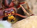
Fig. 18 Frontal oocelles in a hornet

Fig. 19 Pigmented cup eye of the limpet

Fig. 20 Perlboot Nautilus pomilius with pinhole camera eye without lens and cornea

Fig. 21 Jumping spider with large, lens-tipped main eyes and several outer secondary eyes around the head

Fig. 22 Sepia eye with serrated pupil for the simultaneous focusing of the light in two places on the retina

Fig. 23 The scallop has up to a hundred simple eyes


.jpg)


Great diversity of eyes in molluscs
The eyes of molluscs ( mollusca ) evolved seven to eleven times. In this case, a wide diversity of types of eyes, which includes all levels of complexity, from the eye pit of many screws (originated gastropods ), via the hole camera eye of the Nautilus to the highly developed lens eye of cephalopods ( cephalopods ).
The box jellyfish Chironex fleckeri has eight lens eyes, eight slit-shaped eyes and eight lensless pits, 24 eyes in total, which are not connected to a central brain but to a decentralized nervous system. The information processing of the incoming light signals has not been clarified. The eye of the cuttlefish and the octopus is greatly raised from the head, has an overlapping field of view and full all-round vision. The octopus is color blind. Some squids can see colors. The peculiarity of the cuttlefish eye is an oblique, W-shaped pupil , whereby two separate places on the retina come into the light focus (Fig. 22). He can see sharply to the side and to the front at the same time.
The colossal squid has the largest eye in the animal world . With a diameter of up to 30 cm, it is more than the size of a soccer ball. The lens is the size of an orange. The evolutionary necessity for the development of such a large eye, which requires a lot of energy for metabolism, probably does not lie in making it possible to find prey in the dark of great water depth, because swordfish with smaller eyes can do that too . Rather, Nilsson sees the reason as an evolutionary arms race ( coevolution ) between the squid and the sperm whale , their greatest enemies. The sperm whale locates its prey by sound, while the squid with its large eyes at a depth of 500 meters in low light can recognize whales at a distance of up to 120 meters and keep them at a distance.
Compound eyes are present in some mussels ( bivalvia ), and mirror eyes as reflective 'mirrors' evolved in other taxa, for example in the scallop (Fig. 23). The image is also generated in the scallop's eyes (pectuses) by concave mirrors that are placed behind the retina. The lens located directly in front of the retina is used for the optical correction of the strongly distorted mirror image. The mirrors are built on the principle of reflective glass plates. More than 30 layers of the finest guanine crystals are densely stacked, each layer enclosed in a double membrane. Other animals have mirror eyes , including the deep-sea crab gigantocypris , lobsters and lobsters . This form has evidently prevailed where the image quality is less important and the light output is more important. The eyes of molluscs also span a considerable range of sizes. They vary from 20 µm to 27 cm.

The most complex eye in the animal kingdom in the mantis shrimp
One of the most complicated eye constructions in the animal kingdom is that of the mantis shrimp , an order of the arthropod arthropods with around 400 species. Its compound eye, a stem eye, is divided into three parts in its basic structure (Fig. 24). This alone makes it a unique design. The fields of view of the upper and lower sections overlap and are determined by the shape of the eye.
This enables separate spatial vision with each complex eye. In the middle runs a horizontal transverse ligament, which consists of six highly specialized rows of ommatids. With these, the animal can see 100,000 colors, including UV light . To do this, he has sixteen types of color receptors. No animal sees any more colors. In addition, the cancer can perceive light as polarized . The field of view of the ommatid sensor is only 10–15 degrees. Since the two types of eyes, the compound eye and the ommatid ligament, can be moved independently, the cancer can use the compound eye to fix the shape and at the same time recognize and sense details such as color and polarization with the ommatid. The high color visualization as well as the simultaneous polarization ability give various evolutionary explanations: The high color vision ability is explained with sexual selection. Cancer is a strikingly colorful animal. Colors often have a sexual function in the animal kingdom. Mantis shrimp regulate their relationships with other species through showing off and dancing. In addition, the females fluoresce during the mating season. The males recognize the wavelength.
Color recognition and sexual behavior can thus be explained as parallel evolutions. Polarization allows communication with conspecifics or the sexual partner without predators noticing this communication. The evolutionary realization for this requires only a few changes in cells and is easy to select.
Master control genes and gene regulation networks for the eye
In 1995 Walter Gehring discovered the Pax6 gene . It quickly assumed an extremely special position as the master control gene for the evolution and development of all eye types. A master control gene triggers the expression of further genes, which in the case of the eye can be several hundred. Subsequent inductions can also follow, which start again with new master genes and result in a further chain of gene expressions or trigger gene regulation networks.
With Pax6, on the one hand, eye development does not take place completely without expression. On the other hand, ectopic eyes on the leg or antenna of the fruit fly ( Drosophila melanogaster ) can be prodded by Pax6 or its homologous gene Eyeless . Later, the same was partially achieved in vertebrates, including chickens (1995) or using Sox3 in clawed frogs ( Xenopus laevis ) (2000).
In these experiments, ectopic lenses or placodes developed. The fact that the experiments did not lead to such complete results as with the fruit fly suggests that vertebrates are more complex. In any case, vertebrate ocular development ceases entirely if Pax6 is suppressed.
This special position must be reassessed after 20 years. The specialty of Pax6 as a master gene speaks firstly that it is expressed early on the one hand, namely already in eye stem cells, and on the other hand in many tissues during the entire eye development, namely in fruit flies, humans and squid. Eye development is assumed to be independent in these species from different animal phyla. Pax6 can therefore be considered preserved since a common predecessor. Second, reducing its expression leads to decreased eye size in Drosophila , mouse, and humans . Third, Pax6 mis-expression in certain tissues, e.g. B. cause ectopic eyes in the Drosophila wing or leg.
The following facts speak against an outstanding or even sole mastery of Pax6 in eye evolution:
Firstly, the elimination of Pax6 or the homologous gene Eyeless in Drosophila , which also belongs to the Pax6 family, leads not only to the loss of the eye, but also of other parts of the brain, in the extreme case of Drosophila to total head loss .
Second, other genes besides Pax6 play key roles in early eye development, such as the aforementioned Rx1 and Sine oculis (Six), Eyes absent (Eya) or Dachshund (Dach).
These genes can also induce ectopic eyes. Their loss of function also leads to the loss of the eye. They thus show similar master control gene properties as Pax6. The cross-tribal characteristics of Pax6 are put into perspective compared to the capabilities of other master genes today. According to the current state of science, one must speak of the evolutionary conservation of the regulatory network of a whole group of genes which are responsible at the beginning of the chain of evolution of the eye.
Controversy: single or multiple occurrence of the eye
According to the teaching of the evolution theorist Ernst Mayr , which is still widespread today , the eye emerged more than 40 times completely independently of one another.
Recent studies speak of a 50 to 100-fold evolution of the imaging eye.
According to this theory, the vertebrate eye has a different evolutionary origin than about the compound eye of the insect, the hole eye primitive cephalopods or other eye types. If we look more closely, we now see that, on the one hand, the genetic origin of all eye types, the gene regulatory networks , are strikingly the same, which speaks in favor of a homologous , i.e. derived, origin of the eye types.
On the other hand, the “constructions”, i.e. the combined use of all genes involved and the order in which they are activated, and thus also the development paths, differ greatly from one another. Different types of tissue and functional units were created. Thus, the evolution of the eye in Mayr's sense can at least in certain aspects be seen as independent or convergent .
Two different points of view of eye evolution are therefore being discussed today.
On the one hand, there is the view that the eye has a common ancestor, that is, it has a monophyletic origin. Its similar components, for example in vertebrates and insects, are inherited homologously. Above all, the phenotypic results of common gene cascades are homologous according to this view, i.e. they are dependent on a common ancestor. However, this is not mandatory. Even if it is obvious that, for example, similar photoreceptors were created homologously, this does not always have to be the case. The homologous origin of the eye is the view of Gehring and colleagues.
On the other hand, the opinion is also held today that the eye has polyphyletic origins, whereby the similar properties (lens, retina, photoreceptors, etc.) have been acquired independently of one another.
This knowledge has recently become possible on the basis of the analysis of box jellyfish eyes ( Tripedalia cystophora ), a type of jellyfish with 24 eyes around the square umbrella (Medusa). This jellyfish eye has a lens, cornea and retina. The jellyfish are systematically far removed from the vertebrate and its closest ancestors. Despite its cladistic distance from vertebrates, the box jellyfish has a ciliary photoreceptor type in which the visual pigment is embedded in the surface of an eyelash hair, as well as a transmission system for the transmission of light signals comparable to that of vertebrates, which both speaks for homology.
However, the most important crystallines, which are responsible for the optical properties of the lens, are formed in a different way than in vertebrates, i.e. convergent. The question of the single or multiple development of the eye ultimately depends on the definition of the eye and the level of observation. There are clear criteria for the existence of homology in biology. The question can therefore not be answered generally with “homologous” or “convergent”. The example of lens evolution ( Chapter 4 ) most likely represents multiple convergence in eye evolution.
literature
- Walter J. Gehring: Development and evolution of the eyes in: The handicrafts of evolution. Walter J. Gehring tells a genetic theory of development. supposé Berlin 2014
- Georg Glaeser and Hannes F. Paulus: The evolution of the eye. Springer Spectrum 2014
- Simon Ings : The eye. Masterpiece of evolution. Hoffmann and Campe Hamburg 2008
- Trevor D. Lambs: The eye - organ with a past in: Evolution. How she shaped the story of life. Spectrum of Science Special 1/2014.
- Gerald H. Jakobs and Jeremy Nathans: The Strange Color Sense of Primates in: Evolution. How she shaped the story of life. Spectrum of Science Special 1/2014.
Web links
- Africa's light influenced our eye evolution
- David Attenboruough: The evolution of the eye
- Eye evolution (youtube) (differences in vertebrates)
- Richard Dawkins demonstrates the evolution of the eye
- Starfish or sighting star? (Science currently July 5, 2013)
- The evolution of the eye (Richard Dawkins, 2010)
- The New York Academy of Science Magazine: How the Eye evolved (Carl Zimmer), Oct. 2009
- Ancestor of the blue eyes - trend color of evolution
- When evolution is in the eye (Welt.de)
- When evolution is in the eye. From the photoreceptor to the lens eye
- Why the giant squid has eyes the size of a pumpkin
Individual evidence
- ↑ a b c d e Nick Lane: Life. Amazing inventions of evolution . Primus Verlag 2013
- ↑ Andrew Parker: In the Blink of an Eye . Free Press 2003
- ↑ a b c Unique system of photoreceptors in sea urchin tube feet. Esther M. Ullrich-Lüter, Sam Dupont, Enrique Arboleda, Harald Hausen and Maria Ina Arnone. Proceedings of the National Academy of Sciences . PNAS May 2, 2011
- ↑ Charles Darwin: The Origin of Species. Nikol Hamburg 2008, chap. 6th
- ↑ Ramon y Cajal: The retina of the vertebrates. Bergmann 1894
- ^ Gordon Lynn Walls: The Vertebrate Eye and its Adaptive Radiation. The Cranbrook Institute of Science, Bloomington Hills, Michigan; online on openlibrary: The Vertebrate Eye and its Adaptive Radiation by Gordon Walls
- ↑ a b c d e f g Simon Ings: Das Auge '. Masterpiece of evolution. Hoffmann and Campe Hamburg. 2008. Orig .: The Eye: A Natural History , Bloomsbury, 2007, ISBN 978-0-7475-7805-5
- ^ The molecular basis of visual excitation. Les Prix Nobel en 1967, ed. v. R. Granit, Stockholm Nobel Foundation
- ↑ George Wald: The Origins of Optical Activity, Ann. NY Acad. Sci., 1975
- ↑ Nathans J et al. Jeremy Nathans, D. Thomas, DS Hogness: Molecular genetics of human color vision: the genes encoding blue, green, and red pigments . In: Science . 232, No. 4747, April 1986, pp. 193-202. doi : 10.1126 / science.2937147 . PMID 2937147 .
- ↑ Gerald H. Jacobs, Jeremy Nathans: The Strange Color Sense of Primates. In: Spectrum of Science. 5/2010. April 23, 2010, pp. 44–51 , accessed May 23, 2015 .
- ^ Walter Gehring: Historical perspective on the development and evolution of eyes and photoreceptors. The International Journal of Developmental Biology, 48, pp. 707-717, 2004
- ↑ a b Ernst Mayr: That is evolution . Goldmann 2005, p. 250ff
- ↑ a b Michael E. Zuber, Gaia Gestri, Andrea S. Viczian, Giuseppina Barsacchi and William A. Harris. Specification of the vertebrate eye by a network of eye field transcription factors. Development 12/2003, 130, 5155-5167
- ↑ a b M. F. Land, Dan-Erik Nilsson: Animal Eyes , Oxford University Press, Oxford (2002)
- ^ Publications by Nilsson
- ^ A b c Dan-Erik Nilsson: Eye evolution and its functional basis. Visual Neuroscience (2013), 30, 5-20
- ^ A b D. Arendt, K. Tessmar-Raible, H. Snyman, AW Dorresteijn, J. Wittbrodt: Ciliary Photoreceptors with a Vertebrate-Type Opsin in an Invertebrate Brain . In: Science . 306, No. 5697, October 29, 2004, pp. 869-871. doi : 10.1126 / science.1099955 . PMID 15514158 .
- ↑ Trevor D. Lamb, Shaun P. Collin, Edward N. Pugh Jr .: Evolution of the vertebrate eye: opsins, photoreceptors, retina and eye cup . Nature Reviews, 8, 2007, 960-975
- ↑ a b c d e f g h i j Georg Glaeser u. Hannes F. Paulus . The evolution of the eye . Springer Spectrum 2014
- ^ Dan-Erik Nilsson (1996). "Eye ancestry: old genes for new eyes". Current biology: CB 6 (1): 39-42. doi : 10.1016 / S0960-9822 (02) 00417-7 . PMID 8805210
- ^ IR Schwab: You are what you eat . In: BMJ Group (Ed.): British Journal of Ophthalmology . 88, No. 9, September 2004, p. 1113. doi : 10.1136 / bjo.2004.049510 . PMID 15352316 . PMC 1772300 (free full text).
- ^ A b Trevor D. Lamb, Shaun P. Collin, Edward N. Pugh Jr .: Evolution of the vertebrate eye: opsins, photoreceptors, retina and eye cup . Nature Reviews, 8, 2007, 960-975
- ^ RW Scotland: Deep homology: A view from systematics . BioEssays 32 (5): 438-449, (2010) doi : 10.1002 / bies.200900175 . PMID 20394064
- ↑ a b c Fernald, D. Russell: The Evolution of Eyes: Where Do Lenses Come From? Karger Gazette 64: "The Eye in Focus" (2001)
- ^ Dan-Erik Nilsson and Susanne A. Pelger: A pessimistic estimate of the time required for for an eye to evolve . Proceedings of the Royal Society of London. B525, 53-58.1994
- ↑ Richard Dawkins: Summit of the Improbable. Miracle of evolution. Rowohlt 1999. Orig .: Climbing Mount Impropable . Viking Penguin Group London 1996
- ^ W. Westheide, RM Rieger, G. Rieger, G. Rieger (Ed.): Special Zoology. Part 2: vertebrates or skulls. Springer Verlag 2nd edition 2010, page 100. ISBN 978-3-8274-2039-8
- ↑ Johannes W. Rohen, Elke Lütjen-Drecoll: Functional Histology . Schattauer, FK Verlag 4th edition March 2000, p. 476. ISBN 978-3-7945-2044-2
- ^ A. Reichenbach, A. Bringmann (2010). Müller cells in the healthy and diseased retina. New York: Springer. pp 15-20
- ↑ Trevor D. Lambs. The eye - organ with a past in: evolution. How she shaped the story of life. Spectrum of Science Special 1/2014
- ↑ Huxley, Julian (1942/2010): Evolution - The Modern Synthesis. The Definitive Edition, with a Foreword by Massimo Pigliucci and Gerd B. Müller. MIT Press, Cambridge
- ^ Yokoyama, S., and BF Radlwimmer. 2001. The molecular genetics and evolution of red and green color vision in vertebrates. Genetics Society of America. 158: 1697-1710
- ↑ Timothy H. Goldsmith, Birds See the World More Colorful , in Spectrum of Science , January 2007, pp. 96-103; → Spectrum and ( PDF )
- ↑ JK Bowmaker: Evolution of color vision in vertebrates . Eye 12 (3b): 541-547. (1998) doi : 10.1038 / eye.1998.143 . PMID 9775215
- ↑ a b c K. S. Dulai, M. von Dornum, JD Mollon, DM Hunt: The evolution of trichromatic color vision by opsin gene duplication in New World and Old World primates . (1999) Genome Research 9 (7): 629-638. doi : 10.1101 / gr.9.7.629 (currently not available) . PMID 10413401
- ↑ Diana Widermann, Robert A. Barton, and Russell A. Hill. Evolutionary perspectives on sport and competition. In Roberts, SC (2011). Roberts, S. Craig, ed. Applied Evolutionary Psychology. Oxford University Press. doi : 10.1093 / acprof: oso / 9780199586073.001.0001 . ISBN 978-0-19-958607-3
- ^ Nathans, J., and D Thomas (1986). "Molecular genetics of human color vision: the genes encoding blue, green and red pigments". Science 232 (4747): 193-203. doi : 10.1126 / science.2937147 . PMID 2937147 .
- ↑ O.-E. Lund, B. from Barsewisch. The macula in the animal series. Macular diseases. Deutsche Ophthalmologische Gesellschaft Volume 73, 1975, pp 11-17. Jumper
- ↑ L. Spinney: How much of the world do we really see? New Scientist 2265.2000
- ↑ Cottaris, NP, de Valois, RL Temporal dynamics od chromatic tuning in macaque primary vision cortex. Nature, 395, pp. 896-900, 1998
- ↑ L. Spinney. Blind to change . New Scientist, 2265. pp. 28-32
- ↑ DJ Simons, DT Levin: Failure to detect changes to people during real-world interaction . Psychonomic Bulletin and Review 4, p. 644. 1998
- ↑ Paul Simoens: organ of sight, Organum visus. In: Franz-Viktor Salomon, Hans Geyer, Uwe Gille (Ed.): Anatomy for veterinary medicine. 2nd, revised and expanded edition. Enke, Stuttgart a. a. 2008, ISBN 978-3-8304-1075-1 , pp. 579-612.
- ↑ Thomas Geissmann . Comparative primatology. Springer-Verlag, Berlin et al. 2002, ISBN 3-540-43645-6 .
- ^ L. Roth, A. Kleber, Color vision in nocturnal and diurnal geckos . The Lund Vision group, Lund University. 2004
- ↑ Kiama, S G.; JN Maina; J. Bhattacharjee and KD Weyrauch (2001) Functional morphology of the pecten oculi in the nocturnal spotted eagle owl (Bubo bubo africanus), and the diurnal black kite (Milvus migrans) and domestic fowl (Gallus gallus var. Domesticus): a comparative study. Journal of Zoology 254: 521-528
- ^ A. Herrel, JJ Meyers, P. Aerts, KC Nishikawa. The Mechanics of prey prehension in chameleons (2000). Journal of Experimental Biology 203: 3255-3263
- ↑ J.-P. Ewert: Motion perception shapes the visual world of amphibians , In: FR Prete (Ed.): Complex Worlds from Simpler Nervous Systems. Cambridge, MA MIT Press 2004, 117-160
- ↑ Kenneth T. Brown. A linear area centralis extending across the turtle retina and stabilized to the horizon by non-visual cues. Vision Research 10/1969; 9 (9): 1053-62
- ↑ a b c Halder, Georg, Patrick Callaerts, Walter J. Gehring. (1995) Induction of Ectopic Eyes by Targeted Expression of the eyeless Gene in Drosophila. Science. 267: 1788-1792.
- ↑ Michael SY Lee et al .: Modern optics in exceptionally preserved eyes of Early Cambrian arthropods from Australia. In: Nature , Volume 474, 2011, pp. 631-634, doi : 10.1038 / nature10097
- ↑ RD de Voe: Ultraviolet and green receptors in principal eyes of jumping spiders. Journal of Cell Biology , Volume 66, No. 2, pp. 193-207, 1975, doi : 10.1085 / jpg.66.2.193 (currently unavailable) , PMC 2226199 (free full text)
- ↑ Takashi Nagata et al .: Depth perception from image defocus in a jumping spider. Science , Volume 335, No. 6067, 2012, pp. 469-471 doi : 10.1126 / science.1211667
- ↑ Camera eyes in gastropod molluscs , mapoflife.org
- ^ Dan-Erik Nilsson, Eric J. Warrant, Sonke Johnsen, Roger Hanlon and Nadav Shashar: A Unique Advantage for Giant Eyes in Giant Squid . Current Biology 22, 1-6, April 24, 2012. doi : 10.1016 / j.cub.2012.02.031
- ^ Colossal Squid Exhibition. ( Memento from April 20, 2008 in the Internet Archive ) Website of the New Zealand national museum Te Papa on the examined colossal squid.
- ↑ a b Jeanne M. Serb, Douglas J. Eernisse: Charting Evolution's Trajectory: Using Molluscan Eye Diversity to Understand Parallel and Convergent Evolution. In: Evolution: Education and Outreach. 1, 2008, pp. 439-447, doi : 10.1007 / s12052-008-0084-1 .
- ↑ Tsyr-Huei Chiou, Sonja Kleinlogel, Tom Cronin, Roy Caldwell, Birte Loeffler, Afsheen Siddiqi, Alan Goldizen & Justin Marshall: Circular polarization vision in a stomatopod crustacean . In: Current Biology . 18, No. 6, 2008, pp. 429-34. doi : 10.1016 / j.cub.2008.02.066 . PMID 18356053 .
- ↑ C.-C. Chiao, TW Cronin, NJ Marshall: Eye design and color signaling in a stomatopod crustacean Gonodactylus smithii . Brain Behavior and Evolution, 56, pp. 107-122. 2000
- ^ CH Mazel, TW Cronin, RL Caldwell & NJ Marshall: Fluorescent enhancement of signaling in a mantis shrimp . In: Science . 303, No. 5654, 2004, p. 51. doi : 10.1126 / science.1089803 . PMID 14615546 .
- ↑ Bristol University: Mantis shrimps could show us the way to a better DVD, October 25, 2009
- ^ Robert L. Chow, Curtis R. Altmann, Richard A. Lang and Ali Hemmati-Brivanlou. Pax6 induces ectopic eyes in a vertebrate. Development 126, 4213-4222, 1999
- ↑ D. Uwanogho, M. Rex, EJ Cartwright, G. Pearl, C. Healy, PJ Scotting, PT Sharpe. Embryonic expression of the chicken Sox2, Sox3 and Sox11 genes suggests an interactive role in neuronal development Mech. Dev., 49 (1995), pp. 23-36
- ↑ Reinhard W. Köste, Ronald P. Kühnlein, Joachim Wittbrodt, Ectopic Sox3 activity elicits sensory placode formation. Science direct Volume 95, Issues 1-2, July 1, 2000, Pages 175-187
- ↑ Jesper Kronhamn, Erich Frei, Michael Daube, Renjie Jiao, Yandong Shi, Markus Noll, and Åsa Rasmuson-Lestander. Headless flies produced by mutations in the paralogous Pax6 genes eyeless and twin of eyeless. Development 2002 129: 1015-1026
- ↑ MA Serikaku, JE O'Tousa: Sine oculis is a homeobox gene required for Drosophila visual system development . Genetics. 1994 Dec; 138 (4): 1137-50
- ^ Nancy M. Bonini, * Quang T. Bui, Gladys L. Gray-Board and John M. Warrick. The Drosophila eyes absent gene directs ectopic eye formation in a pathway conserved between flies and vertebrates. Development 124, 4819-4826 (1997)
- ↑ TA Heanue, RJ Davis, DH Rowitch, A. Kispert, AP McMahon, G. Mardon, CJ Tabin Dach1, a vertebrate homologue of Drosophila dachshund, is expressed in the developing eye and ear of both chick and mouse and is regulated independently of Pax and Eya genes . Mech Dev. 2002 Feb; 111 (1-2): 75-87
- ↑ Z. Kozmik, Michael Daube, Erich Frei, Barbara Norman, Lidia Kos, Larry J. Dishaw, Markus Noll, Joram Piatigorsky: Role of Pax Genes in Eye Evolution A Cnidarian PaxB Gene Uniting Pax2 and Pax6 Functions . (2003) Developmental Cell 5 (5): 773-785. doi : 10.1016 / S1534-5807 (03) 00325-3 . PMID 14602077
- ↑ a b Joram Piatigorsky: A Genetic Perspective on Eye Evolution: Gene Sharing, Convergence and Parallelism . Evolution: Education and Outreach 1 (4): 403-414 (2008)
- ^ A b Luitfried Salvini-Plawen : Photoreception and the Polyphyletic Evolution of Photoreceptors (with Special Reference to Mollusca) , American Malacological Bulletin 01/2009; doi : 10.4003 / 006.026.0209 .
- ^ WJ Gehring: New perspectives on eye development and the evolution of eyes and photoreceptors. J Hered. 2005 May-Jun; 96 (3): 171-84. Epub 2005 Jan 13
- ↑ Multi-eyed jellyfish helps with Darwin's puzzle . Newscientist.com (May 14, 2005)