Nerve cell
A nerve cell , also neuron (from ancient Greek νεῦρον Neuron , German , tendon ' , tendon';, nerve '), is one on impulse conduction and synaptic transmission specialized cell , as cell type in tissue animals and in almost all multicellular animals occurs. The totality of all nerve cells of an animal together with the glial cells form the nervous system .

The pyramidal cell with large dendritic tree in the center expressed here green fluorescent protein . GABA -producing interneurons can be seen in red .
(Length of the rule below right: 100 µm )

(drawn by Santiago Ramón y Cajal , 1899)

A typical mammalian nerve cell has a cell body and two types of cell processes: the dendrites and the neurites or axons . The ramified dendrites primarily take up excitation from other cells. The neurite of a neuron, which is enveloped by glial cells, can be over a meter long and is initially used to transmit an excitation of this cell to the vicinity of other cells. A voltage change is transmitted via the extension by allowing brief ion currents through special channels in the cell membrane .
The axon ends are in contact with other nerve cells, muscle cells ( neuromuscular endplate ) or with gland cells via synapses , where the excitation is rarely passed on directly electrically, but is mostly transmitted chemically by means of messenger substances ( neurotransmitters ) . Some nerve cells can also release signal substances into the bloodstream, e.g. B. modified neurons in the adrenal medulla or in the hypothalamus as secretion of neurohormones .
It is estimated that the human brain, with a mass of one and a half kilograms, consists of almost ninety billion nerve cells and a similar number of glial cells.
The nerve cell is the structural and functional basic unit of the nervous system. Its designation as a neuron goes back to Heinrich Wilhelm Waldeyer (1881).
construction
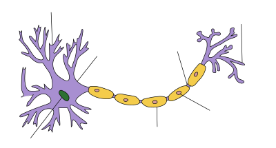
| Structure of a nerve cell |
|---|

The cell body
Every nerve cell has a body that, like other cells, is called a soma or, in neuroanatomical terms, a perikaryon. The perikaryon here comprises the plasmatic area around the cell nucleus , without cell processes such as neurite and dendrites. In addition to the cell nucleus, the cell body contains various organelles such as rough and smooth endoplasmic reticulum , mitochondria , the Golgi apparatus and others. Characteristic of the cell body of neurons is the condensed appearance of the endoplasmic reticulum and its accumulation as Nissl substance to form Nissl clods, which, on the other hand, are absent in the processes and also in the axon hillock. All proteins and other important substances that are necessary for the functioning of the nerve cell are formed in the soma ; Depending on the type and size of the neuron, its perikaryon measures between 5 µm and more than 100 µm.
Excitations from other cells are transmitted locally to the branched dendrites and spread as electrical voltage changes across the membrane of the nerve cell, becoming weaker with increasing distance. Overlaying each other, they converge in the area of the perikaryon, where they are collected and processed in an integrating manner. This is done through spatial and temporal summation of the various changes in the membrane potential .
It depends on the result of the summation at the location of the axon mound whether the threshold potential is exceeded and thus an action potential is now formed or not (see all-or-nothing law ). The resulting action potentials are an expression of the excitation of a nerve cell. You are successively over the Axonfortsatz forwarded . At its endings excitation is to other cells transmitted .
The dendrites
Various plasmatic processes extend from the cell body of a nerve cell . The dendrites (Greek δένδρον dendron , German 'tree' ) are finely branched nerve cell processes that grow from the soma and form contact points for other cells, the excitation of which can be transferred to the nerve cell here. The neuron is linked to a specific cell via a synapse and picks up signals with the locally assigned postsynaptic membrane region of a dendrite. The dendrite tree of a single nerve cell can have several thousand such synaptic contacts through which various signals flow to it, each of which is mapped locally as certain changes in the postsynaptic membrane potential. The individual contact points can be designed differently; some neurons have special formations in the form of dendritic thorns . Synaptic activation on the dendrite alone can bring about changes that can last for a long time (see synaptic plasticity ).
The axon mound
A special area of the cell body free of Nissl clods is formed by the cone of origin of the neurite or axon hill, from which one axon of a nerve cell emerges. Here the threshold potential is significantly lower, so that postsynaptic potentials are most likely to trigger an action potential at this point in the perikaryon. The action potentials formed in the subsequent first section of the axon, its initial segment, are passed on via the axon. The axon mound is the place where postsynaptic changes in potential are integrated and converted into series of action potentials, thus recoding analog signals into digital signals. Due to its low threshold potential and the location of the axon hill, it is ensured that, if the nerve cell is excited, action potentials arise at this point and are passed on via its axon.
The axon
The axon (Greek ἄξων axon , German , axis' ) of a nerve cell is the springing on the axon hillock gliaumhüllte neurite over which their excitation to other cells forwarded is. Action potentials triggered in the initial section are passed on via the axon and its side branches (collaterals) to the terminal sections, which usually form an end button as a presynaptic terminal . Depending on the location of the target cell and depending on the type and size of the nerve cell, there are considerable differences in the length and diameter of the axons. The axons of the nerve cells of mammals are about 0.05 µm to 20 µm thick and in humans between 1 µm and 1 m long.
In the course of the process, these nerve cell extensions are enveloped by glial cells of the nervous system - in the peripheral by the Schwann cells and in the central by the oligodendroglia . The axon and sheath together form a nerve fiber. When glial cells envelop the axial cylinder with multiple wraps, their membrane lamellae form an insulating medullary or myelin sheath around the axon, with a narrow gap between the individual glial cells at the cell boundaries - called Ranvier's nodules following sections (internodes). This structure characterizes myelinated nerve fibers and enables faster, leaps and bounds conduction of excitation than with the so-called unmarked nerve fibers with a simple covering without a myelin sheath.
In the cytoplasm of the axon (axoplasm) there is a particularly structured cytoskeleton made up of neurofibrils and microtubules , which is used in particular for axonal transport within this, often extremely long, cell process. In this way, the proteins and membrane sheaths synthesized in the soma are transported to the terminal axon ( anterograde ). But there is also rapid transport of substances in the opposite direction towards the cell body ( retrograde transport ).
The myelin sheath
The enveloping of an axon by glial cells with multiple, isolating wrappings in sections is called the myelin sheath (Greek μυελός myelos , German 'marrow' ). Such medullary nerve fibers conduct signals about ten times faster than medullary axons of the same diameter and thus enable an organism to react faster to stimuli from the environment. While oligodendrocytes myelinate axons in the central nervous system , it is Schwann cells in the peripheral course that envelop an axon and wrap around it up to fifty times for the myelin sheath. Since the biomembranes of the two types of glial cells differ somewhat, the myelin peripherally has a different composition of phospholipids and proteins than the central one.
A single Schwann cell, wrapped around it in tightly packed layers, consisting almost entirely of lamellae of its cell membrane, forms an approximately 1 mm long myelin sheath section for the axon located inside. A myelinated axon segment ( internode ) is separated from the next at the node (nodus). The myelin sheath is thus formed lengthways from a row of glial cells and interrupted between adjacent ones at regular intervals by narrow so-called Ranvier rings . These play an essential role in the transmission of action potentials along the myelinated axon. Because here the voltage changes jump from ring to ring ( saltatory excitation conduction ) in a depolarizing manner because of the insulating sheath , where an action potential is then built up, while with non-myelinated nerve fibers this occurs progressively along the entire length of the axon membrane ( axolemm ) and thus lasts longer. The initial and terminal parts of an axon are usually not myelinated.
The end button
An axon and each of its branches into collaterals ends with a so-called end button , also called an end bulb or end plate or axon terminal , which represents the presynaptic part of a synapse . Synapses link nerve cells with one another or with other cells in such a way that an excitation can be transmitted to individual downstream cells.
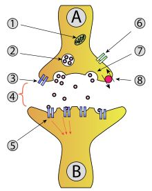
The end button on the terminal axon serves as a presynapse of the "transmitter cell". In the area under the presynaptic cell membrane, it contains molecules of a neurotransmitter packaged in separate membrane sheaths of synaptic vesicles ( synaptic vesicles ) . Achieved at a continuous excitation propagated action potential the Endköpfchen, so flow through voltage-dependent calcium channels increased calcium - ion (Ca 2+ ) in the presynaptic terminal. This short-term calcium influx triggers a chain of interactions between special proteins, as a result of which synaptic vesicles are attached to the presynaptic cell membrane and fuse here by membrane fusion. The vesicle content with neurotransmitter is released into the narrow intercellular space by exocytosis .
On the opposite side of the synaptic gap is as postsynapse the postsynaptic membrane area of the "recipient cell" that here with receptors is equipped to which bind the neurotransmitter molecules. Ion channels are then opened, which leads to a change in electrical voltage on the postsynaptic membrane, which is then spread to neighboring regions. The synaptic gap between the two cells is an approximately 30 nm narrow gap and part of the extracellular space in which the transmitter molecules are distributed by diffusion .
The synapse

1 mitochondrion , 2 synaptic vesicles , 3 autoreceptors , 4 synaptic clefts with released unbound neurotransmitters , 5 postsynaptic receptors , 6 calcium channels , 7 exocytosis of the vesicle, 8 active transport across the cell membrane
function
The synapse is the place where excitation can be transferred from one cell to another . A neurotransmitter is usually used to bridge the narrow gap between the cells, known as the synaptic gap . Synapses can be understood as interfaces between cells through which nerve cells communicate with other cells.
A single nerve cell can be related to other cells via numerous synapses, with regard to both incoming and outgoing signals. A Purkinje cell in the cerebellum, for example, receives input signals from other neurons via around 100,000 dendritic synapses. A granule cell in the cerebellum sends output signals to other neurons, including Purkinje cells, via several hundred synapses. The total number of synapses in the human brain is estimated to be a little under a quadrillion .
The neurons as parts of the nervous system are rarely directly connected to one another by electrical synapses , but mostly via chemical synapses . This transmission of signals by means of messenger substances can be modified or modulated in various ways . The conditions for signal transmission are therefore not completely fixed, but can be shaped within certain limits, which is known as synaptic plasticity . Linked in this way, neurons form a malleable neural network , which can change in its effect through the act of use ( neural plasticity ).
In terms of information technology, the basics of the networking of neurons can be reproduced in a greatly simplified manner. An artificial neural network with a different architecture can also be designed, which can be trained step by step, for example, so that it is suitable for recognizing complex patterns. Here, the learning process is anchored in shifts in the weightings between neuron elements or their threshold value.
Neurotransmitters
Neurotransmitters are used at chemical synapses as messenger substances for the transfer of excitation from one nerve cell to another cell ( transmission ). The transmitters are released presynaptically when the "sender cell" is excited, bridge the synaptic gap between the cells and are received postsynaptically by the "recipient cell " using special receptor proteins . These recognize the respective transmitter molecule specifically by its spatial shape and charge distribution through complementary structures and bind it reversibly.
The binding leads to a remodeling of the receptor structure (conformational change). This can directly ( ionotropically ) trigger or indirectly ( metabotropically ) influence the opening of ion channels in the membrane, thereby allowing brief ion currents and thus temporarily changing the membrane potential of this region. Depending on the configuration of the postsynaptic membrane, a potential difference (postsynaptic potential, PSP) is produced as a local response, which either favors or triggers the excitation of the recipient cell (exciting, excitatory PSP ) or makes it difficult or impossible for a short period of time (inhibiting) to excite it , inhibitory PSP ).
The excitation transmitted by means of the transmitter has either an exciting or an inhibitory effect on the downstream cell at a chemical synapse, which is determined by the types of receptors and the types of ions of the affected membrane channels in the respective linked cell. In neuromuscular synapses, for example, impulses from excited nerve cells at motor end plates are transmitted to muscle fibers using acetylcholine (ACh) as a transmitter substance and have a stimulating effect on these effectors , so the postsynaptic end plate potential can trigger an action potential in these cells. This leads to contraction in the muscle cells , which can be seen as a shortening of the muscle in the associated successor organ . The effect of the possibly rebinding transmitter is limited here primarily by its enzymatic breakdown ( acetylcholinesterase ) in the synaptic gap.
Working method

A nerve cell receives a signal in that neurotransmitters , for example released by an upstream cell, bind to special receptors in the postsynaptic membrane in the dendrites or in the soma of the cell to be excited. If the excitation is transmitted in this way, it is passed on via the dendrites to the soma of the nerve cell and from there to the axon mound. Each of the incoming depolarizations at the various synapses of the nerve cell changes the membrane potential on the axonal membrane, where an action potential is triggered when a threshold value is exceeded. In general, the closer a synapse attaches to the soma, the stronger its influence on the nerve cell, the longer the path that the excitation has to cover, the weaker the influence. A stronger stimulation of a dendrite thus results in a stronger depolarization (see second graph in the picture on the right). Almost simultaneously incoming stimuli add up in their effect, which means that an excitation potential builds up within the cell and on the axon hill (summation).
In the axon mound, certain factors now decide according to the rules of the all-or-nothing law about the triggering of an action potential , whereby it is decided whether the threshold potential has been reached and exceeded. If this is the case, the action potential is released along the axon through the depolarization of the axon. This in turn happens on the biomembrane , the so-called axolemm , which separates the intracellular area (inside) from the extracellular area (outside).
Ions are located inside the axon as well as outside the membrane . The axon's biomembrane causes different concentrations of ions to exist between the inside and outside , so that a different electrical charge is applied to the axon's outer wall than the inside - the inside of the cell is negatively charged. One speaks of a polarization or a resting potential . The axolemm creates and maintains polarization with the help of a sodium-potassium pump , named after the ions of the elements sodium and potassium , which play an important role in the transmission of excitation. This process is called active transport , because it requires energy to be supplied. If an action potential moves due to the change in the concentration gradient of the ions within the axon along the axon to the terminal button , this electrical impulse reaches a limit at the end of the axon, since the electrical signal cannot be transmitted through the synaptic gap between the two cells is. The stimulus is chemically passed on via the synapses and transferred to another cell in the same way as the process already described.
As soon as an action potential has been triggered, the cell needs time to rebuild the membrane potential ( repolarization ). During this pause, also known as the refractory phase , no new action potential can be triggered. So if one of the stimuli coming in one after the other is so strong that the cell forms an action potential and the subsequent stimulus comes in during the refractory period, the cell does not develop a new action potential for it.
The more action potentials the cell fires per unit of time, the stronger the stimulus. The excitation conduction is basically possible in both directions. Due to the inactivation of the sodium channels, the action potentials are forwarded preferably in one direction. It is also said that the nerve cell fires. It can do this up to 500 times in one second.
The prerequisite for the functioning of the neuron is therefore its ability to receive and transmit an electrical impulse. Important factors play a role: the electrical excitability (receiving the impulse), the resting potential (the possibility of integrating it), the action potential (forwarding and transmitting it) and the conduction of excitation (transmitting it in a targeted manner).
Differentiation of nerve cells in terms of structure and function
Morphological distinction

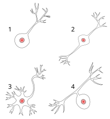
1 unipolar nerve cell
2 bipolar nerve cell
3 multipolar nerve cell
4 pseudounipolar nerve cell
The neurons found in the nervous system can differ in structure and function in several ways. Visually, they can be easily classified by the type and number of their appendages.
Unipolar nerve cells
There are unipolar nerve cells that are equipped with only a single, short process, which usually corresponds to the neurite or axon . They are found, for example, as primary sensory cells in the retina of the eye .
Bipolar nerve cells
A bipolar nerve cell is a neuron with two processes. Bipolar cells are specialized sensor neurons that mediate certain senses. As such, they are part of the sensory transmission of information for the sense of smell, sight, taste, touch, hearing and balance.
The bipolar cells of the retina and the ganglia of the auditory balance nerve are usually given as examples . In the absence of specific information, the term usually refers to the cells that make up the retina.
Multipolar nerve cells
A very common group are multipolar nerve cells . They have numerous dendrites and an axon. This cell type can be found, for example, as motor nerve cells in the spinal cord .
Pseudounipolar nerve cells
The pseudounipolar nerve cells also have two processes . There, however, the dendrite and axon merge near the cell body. They are found in sensitive nerve cells whose perikarya are located in the spinal ganglia . The excitation does not first pass through the perikaryon, but passes directly from the dendritic to the neuritic axon.
Differentiation according to myelination
Another possibility for visual differentiation is the development of the myelin sheath by Schwann cells in the area of the axon. Both a myelinated and a myelinated form exist here, whereby those nerve fibers are referred to as myelinated, the axons of which are surrounded by a strong myelin sheath. If this myelin sheath is very thin, the nerve fiber in question is referred to as having little or no myelin (with differentiated cells).
Functional distinction
The individual functions of the nerve cells offer another possibility of differentiation. A general distinction is made here between motor neurons, sensory neurons and interneurons .
- Sensory neurons, also known as afferent nerve cells , transmit information via nerves or nerve fibers from the receptors of the sensory organs or from various organs to the brain and spinal cord or to the nerve centers of the intestine . The information transmitted is used for perception and motor coordination .
- Motor neurons, also called efferent neurons or motor neurons called that transmit impulses from the brain and spinal cord to the muscles or glands where they trigger such as a contraction of the muscle cells , or provide for the secretion of secretions or the secretion of hormones .
- Interneurons make up the largest number of neurons in the nervous system and are not specifically sensory or motor. They process information in local (local) circuits , or convey signals over long distances between different parts of the body. They act as an intermediary. A distinction is made here between local, regional, segmental and intersegmental interneurons.
Pathology of the nerve cell
Pigment deposits
In certain nuclei of the central nervous system, deposits of pigment are normally observed within the nerve cells. Particularly noticeable is the neuromelanin in the substantia nigra and the locus caeruleus , which gives the neurons a characteristic brown-black appearance and allows these core areas to be recognized with the naked eye. The proportion of yellowish lipofuscin increases with age and is particularly observed in the dentate nucleus of the cerebellum and the lower nucleus of the olive . In certain dementia diseases, such as Alzheimer's disease , characteristic eosinophilic inclusion bodies of the nerve cells are observed.
Effects of poisons
Nerve toxins usually act on the existing protein structures of the cell and in this way disrupt the exchange of information between the neurons. There are numerous examples of such neurotoxins, one of which is diisopropyl fluorophosphate (DFP). If DFP gets into the body, it binds irreversibly to the enzyme acetylcholinesterase , which is responsible for the breakdown of acetylcholine in the synapse of, for example, motor neurons . This increases the concentration of the acetylcholine transmitter in the synaptic gap and the innervated muscle cells are permanently excited. The subsequent overexcitation can lead to severe cramps in the affected organism and even to fatal outcome (death).
Tetrodotoxin (TTX, poison of the puffer fish ) blocks sodium channels. Tetraethylammonium (TEA) blocks potassium channels.
- Some known poisons are
| Poison | Occurrence |
|---|---|
| Alkyl phosphates | Plant poisons , war gases |
| Formic acid | Nettles |
| Hyoscyamine | Deadly nightshade |
| Botulinum toxin | spoiled food |
| Curare | Plant poison |
| nicotine | plants |
| Muscarin | Mushroom poisons |
Examples of specialized nerve cell types
| Illustration | Surname | localization |
|---|---|---|
 |
Ganglion cells | retina |
| Motor neurons | Spinal cord , muscles | |
 |
Pyramidal cells | Cortex , hippocampus |
| Granule cells | Cortex , hippocampus , cerebellum , olfactory bulb | |
 |
Purkinje cell | Cerebellum |
| Olfactory cells | Epithelium of the nasal mucosa |
See also
- Neuron theory
- Neuropil
- Neurogenesis
- Neuroprosthesis
- History of brain research
- Developmental Neurobiology
- smRNA
literature
- Robert F. Schmidt , Gerhard Thews , Florian Lang (eds.): Physiology of humans (= Springer textbook ). 28th edition. Springer, Berlin 2000, ISBN 3-540-66733-4 , pp. 199-206 .
Web links
- Structure and explanation
- The construction of a neuron
- The nerve cell
- Brain cells (English)
- When neurons communicate Telepolis
- Interactive web presentation of neural processes
- electron microscope images of ganglion cells
- Manfred Spitzer : Neurons grow ( Memento from March 3, 2013 in the Internet Archive ). Video from the BR-alpha series "Mind and Brain" (approx. 15 minutes)
Notes and individual references
- ↑ In the human body, the longest neurites are found in motor neurons in the anterior horn of the spinal cord, which conduct impulses from there to the muscles of the distal lower extremity, for example from the spinal cord segment S1 to muscle fibers of the short flexor of the big toe . Nerve fibers innervating this muscle run in nerves of the peripheral nervous system (via sacral spinal nerves to the lumbosacral plexus , then in the sciatic nerve (sciatic nerve), after it bifurcates in the tibial nerve and after its branching then in the medial plantar nerve , if they are muscle fibers of the medial head of the short big toe flexor). Nerve cell extensions of approximately the same length are found in pathways of the central nervous system as neurites of pyramidal cells in the cortex of the cerebrum, which run from there via pyramidal pathways to spinal cord sections and end, for example, in sacral segment S2.
- ↑ According to the results of Azevedo and Team 2009 ( PMID 19226510 ), the number of nerve cells in the brain of an adult male is estimated to be around 86 ± 8 billion, that of glial cells to around 85 ± 10 billion; the degree of precision of the method used - counting immunohistochemically labeled NeuN (+) or NeuN (-) cells from fractionated tissue samples ( isotropic fractionator ) - can not yet be reliably stated according to Lyck and Team 2009 ( PMID 19520115 ), it seems, according to Bahney and V. Barthfeld 2014, however, at least valid for glial cells.
- ↑ Welsch: Textbook Histology. 2nd edition, Elsevier, Munich 2006.


