History of brain research
The history of brain research goes back to the first knowledge of brain anatomy in prehistoric times. The insight that the brain is the seat of cognitive abilities can be demonstrated for the first time in ancient Greece , but its functioning remained largely unknown until the end of the Middle Ages . While the structure of the brain was examined more closely as early as the Renaissance after the resurgence of autopsies , methods for gaining experimental knowledge about its function have only been available since the 18th century . Most of the current state of knowledge on brain anatomy and neurophysiology was acquired around the middle of the 19th century through systematic research on animals and observations on sick and injured people. In addition, non-invasive methods have been available since the middle of the 20th century , which have contributed to many other findings through experiments on healthy test subjects.
prehistory

Based on finds from early Egypt , we know that 5000 years ago people began their first surgical interventions in the central nervous system, as can be seen from systematic cranial openings ( trepanations ) on skulls from this time. About 70 percent of skulls that show such features show signs of healing and therefore suggest that the patient survived the procedure by months or even years. This can be seen as the birth of neurosurgery . Neolithic trepanations are also known from all over Europe and Latin America.
The Egyptians' anatomical knowledge of the brain emerges from the surprisingly systematic and rational papyrus Edwin Smith . This papyrus, which was written in Egypt in 1550 BC, probably goes back to writings that already existed around 3000 BC and are considered the oldest medical documents in human history. The papyrus describes the brain, its organization in the gyri and sulci, the spinal cord , the meninges and the surrounding bones. The blood vessels, tendons and nerves are still indiscriminately referred to as "channels". The Egyptians were also not yet clear about the function of the brain: although they had learned that severe head injuries could result in loss of language, the heart was considered to be the seat of the soul and all mental faculties. While it was not allowed to be touched during embalming and other organs such as lungs , liver and stomach were given to the corpse for its afterlife, the Egyptians removed the brains of their dead without hesitation.
Antiquity

Around 500 BC Alkmaion of Croton is said to have been the first to discover the optic nerves and other sensory nerves. Alkmaion developed the idea that nerves are hollow and enclose a medium ( kenon ) that guides the sensory impression to the brain. Although autopsies on humans were unthinkable for religious reasons at that time, the Corpus Hippocraticum collection attributed to Hippocrates of Kos (approx. 460-370 BC) already clearly identified the brain as the seat of sensation and intelligence and recognized that the brain was previously Epilepsy, considered “sacred”, is a disease of the brain. The first autopsies were possible at the time of Herophilos of Chalcedon (around 325-255 BC). He correctly described the rough anatomy of the brain, but assumed that human intelligence or soul forces were not located in the brain tissue, but in three cerebral ventricles , the fluid-filled chambers of the brain, which he distinguished for the first time . Erasistratos (around 305–250 BC) distinguished between motor and sensory nerves, recognized four cerebral ventricles (based on the division of the first into a right and left ventricle), localized the soul in the convolutions or in the meninges and undertook neurophysiological experiments such as B. Brain slices and artificial lesions . For a long time, however, the new discoveries could not completely displace the older idea that sensation and understanding are assigned to the heart. Its best-known representative was Aristotle (384–322 BC), who viewed the brain as a cooling organ.
Galen's Ventricular Doctrine (around 177 AD):
In the post-Christian period, the Greek doctor Galen (around 129–216 AD) carried out careful studies of animal anatomy and explained the function of individual nerve tracts using numerous vivisections . He contradicted the Aristotelian view of the brain as a cooling organ. Galen also described the sympathetic nervous system for the first time , but did not yet correctly understand its function. Autopsies of people were banned in Rome , where the main focus of his work was. However, Galen had the opportunity to examine wounded gladiators, and apart from that, applied much of his findings from animal studies to humans. In his investigations of the brain, following Herophilus, he mainly concentrated on the liquor- filled brain chambers. Among other things, he studied how cuts and pressure affect them. Galen believed that there was a substance in the ventricles which he called pneuma psychikon (Latin spiritus animalis ) and which was able to transport sensory perceptions to the brain through the nerves, which were imagined to be hollow, but also to activate muscles. He claimed that this pneuma psychikon is obtained, among other things, from the rete mirabile , a fine network of blood vessels that is located at the base of the brain in sheep. The fact that the human brain has no corresponding structure at all remained undiscovered for around 1300 years.
Galen regarded the ventricles essentially as the reservoir of the pneumas (not of the soul, which, like Erasistratos, he assumed to be in the brain matter) and admitted the brain matter to an important role in cognitive processes; this is how he was the first to describe age-related brain wasting . Christian church fathers soon developed a synthesis of Galen's medical knowledge with the theologically motivated human image of Christianity. In the course of this development, the ventricles became the focus of attention - they were now considered to be the seat of the immortal soul until the end of the 18th century (for example by Samuel Thomas von Soemmerring ). In addition, the various ventricles were now assigned specific functions. An early representative of this doctrine of localization was Bishop Nemesius of Emesa (around 400). In this modified form, Galen's teaching was elevated to a dogma and dominated the concept of man well into the 18th century.
middle Ages
The knowledge of Western European medicine and thus also of brain research fell behind the level of antiquity in the Middle Ages. The little research in Europe concentrated on monastic herbal studies, which had little to contribute to brain research. The only thing worth mentioning is Albertus Magnus , who continued the theory of the ventricles around 1250. According to him, the spiritus animalis flows like a Roman fountain from one ventricle to the next and thus mediates the process from perception through thinking to memory.
In the meantime, medical research was continued in the Byzantine and Arabic cultures, so that Arabic medicine dominated the knowledge of brain research until the Renaissance. Around 900 Rhazes (Abu Bakr Mohammad Ibn Zakariya al-Razi) examined the brain anatomically more precisely and described seven of the twelve cranial nerves and 31 of the spinal nerves arising from the spinal cord in his work Kitab al-Hawi Fi Al Tibb (Arabic: Secret of Secrets) . Already 100 years later, Abu l-Qasim az-Zahrawi (also known as Abulcasis or Albucasis) was already describing surgical interventions to heal neurological diseases of the central nervous system. However, findings in Persian-Arabic medicine were by no means limited to the central nervous system; they also made functional assumptions about the peripheral nervous system. Thus, at the turn of the millennium , Abu Ali al-Hasan Ibn al-Haitham (Alhazen) compared the functioning of the eye with a device similar to a camera. Even Abu Ali al-Husain ibn Abd Allah ibn Sīnā (Avicenna) described the eye and the principles of vision in his Qanon ( Canon of Medicine ) and Abu Ruh Muhammad ibn Mansur ibn abi 'Abdallah ibn Mansur al-Yamani (also known as al- Jurjani and Zarrin-Dast described several surgical procedures on the eye in his book Nur al-ayun (Light of the Eye) in 1088. However, it is unclear how often such operations were actually carried out in practice.
Renaissance
During the Renaissance , the first new impulses for brain research came from Italy, where it gradually became possible again to dissect human corpses. This work was initially performed by barbers while the anatomist watched from a raised seat and quoted from textbooks. The doctor Mondino dei Luzzi (around 1275-1326) wrote Anathomia Mundini (1316), the first work on anatomical sections since the beginning of the Middle Ages. Leonardo da Vinci (1452–1519) made significant contributions to a more realistic graphic representation of anatomical structures. In 1490, after numerous sections , he was the first to depict a sagittal brain slice in a manuscript that was later called Codex Windsor , but initially kept the drawing a secret. In 1504 he also made wax casts of the ventricles of an ox's brain in order to study their shape more precisely. The German anatomist Johann Dryander (1500–1560) drew the first axial brain slice in 1536 , but the perspective was not yet correct.
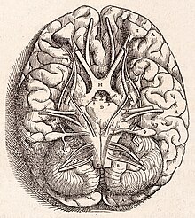
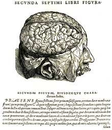
Galen's authority, which had been absolute in the Middle Ages, has been questioned since the 16th century: The doctor Jacopo Berengario da Carpi (around 1470–1530) mentioned in his De Fractura calve sive cranei (1518) that he was not in was able to find the rete mirabile in humans. The Flame Andreas Vesalius (1514–1564) finally dared to break openly with Galen's tradition. He carried out extremely precise anatomical research in Padua for the time and with his works Tabulae Anatomica and De humani corporis fabrica foundations of (neuro-) anatomical research put. Vesalius expressed deep respect for Galen's conscientious research, but had recognized that he had transferred numerous findings from studies of animals to humans and had sometimes come to incorrect statements. Vesalius made brain slices that were used as a reference for two centuries and already distinguished between gray and white matter of the cerebral cortex. He doubted the localization of brain functions in the ventricles and found that even very thick nerves, even on closer examination, did not reveal any cavity that could have been used to carry pneuma. Finally, Vesalius described the pineal gland and pons , which at that time were the only known structures of the brain that did not occur twice ( i.e. once in each hemisphere ). Vesalius' new findings were initially only accepted by part of the professional world. In particular, his Paris teacher Jacobus Sylvius (1478–1555) used harsh words to oppose any criticism of Galen's work.
About 20 years after Vesalius' works, the clearly detailed, but also more specialized work De auditus organis by Bartolomeo Eustachi appeared in 1564 , which for the first time provided information about the structure and possible functioning of the acoustic sensory apparatus. The Eustachian tube , which connects the ear and mouth, is named after Eustachi . A number of other works testify to the active neuroanatomy research in Italy: Gabriele Falloppio described some of the cranial nerves, but could show little new knowledge compared to the work of Rhazes. In 1564, in a work by Giulio Cesare Aranzi , the name hippocampus was used for the first time , and Constanzo Varolio named the bridge already described by Vesalius ( Pons Varolii ) in 1573 .
17th century

At the beginning of the 17th century, the English doctor William Harvey (1578–1657) caused a sensation with the discovery of the blood circulation . Previously it was assumed that blood was constantly being produced and used in the organs. The French philosopher René Descartes (1596–1650) realized that the heart was not much more than a mechanical pump, to the assertion that the brain is also constructed like a very complex machine. Consequently, he denied animals any ability to feel and think. Human reflexes and vegetative functions are also to be understood purely mechanically, only feelings, conscious perceptions, reflections and volitional actions are the result of an immortal and immaterial soul , which he imagines interacts with the body in the pineal gland . Descartes had no medical training, but occasionally dissected animal heads and organs, which he obtained from the local slaughterhouse. Many of his assumptions about the functioning of the brain were already considered by contemporary anatomists as speculative and implausible; So the theory emerged that the pineal gland controls the movements of the pneumas in the ventricles, as early as 300 BC. At Herophilos and has already been rejected by Galen. Nevertheless, Descartes' philosophy had a lasting influence on brain research: He is considered the spiritual father of dualism , which postulates a dichotomy of all beings into matter and spirit and to this day not only corresponds to the understanding of many laypeople, but has also shaped the ideas of numerous researchers.

In the middle of the 17th century, a group of naturalists formed in Oxford who called themselves “virtuosi” and actively discussed neuroanatomical questions. Its members included Richard Lower , Robert Boyle and Christopher Wren , among others . In this inspiring environment, the doctor Thomas Willis (1621–1675) dissected a large number of different animals in addition to humans and published his work Cerebri anatome in 1664 . Incorporating realistic drawings by Wren, it immediately became the standard work on the anatomy of the nervous system and cerebral blood vessels . While Descartes suspected that the mental functions were still located in the ventricles, Willis followed Vesalius' considerations and now finally moved the brain matter into the center of attention. He held the gray matter responsible for the production of the pneumas, the white matter for its forwarding; While he assigned the cerebrum a function in conscious movement and thinking, he held the cerebellum responsible for the control of organs and unconscious movements. Willis coined numerous technical terms, including “ neurology, ” and regarded many mental illnesses that were treated by priests rather than doctors during the Middle Ages as organic diseases of the brain.
At the same time, the Italian Giovanni Alfonso Borelli (1608–1679) questioned the existence of a gaseous spiritus animalis for the first time after he had cut the nerve of an animal submerged under water and no ascending gas bubbles could be observed. Instead, he suspected the existence of a fluid, the succus nerveus , which was pressed into the extremities by the hollow nerves and thus should induce the actions according to pneumatic principles. Other brain anatomical discoveries of that time go back to the German Franciscus de la Boe Sylvius , who described the large lateral fissure on the brain surface and examined the connection between the third and fourth ventricles. Both structures are still named after him as Fissura Sylvii and Aquaeductus Sylvii .
18th century
Nerves as electrical conductors
The first compound light microscopes appeared as early as the end of the 16th century . At the end of the 17th century, the Dutchman Antoni van Leeuwenhoek (1632–1723) developed it much further and pointed out as early as 1674 that he had not succeeded in using this device to discover the cavity in the optic nerve ( optic nerve ) of a cow it should contain according to the anatomical understanding of the time. Although microscopes still suffered from limited magnification and severe distortion in the 18th century, they made it possible for the Italian Felice Fontana (1730–1805) to describe the axons of the nerves precisely for the first time.
Also around 1600 the English physician and physicist William Gilbert (1544-1603) began scientific research into electricity . Electric eels and electric rays had already been used therapeutically in antiquity, but their effects on humans were not clarified until the 18th century: John Walsh (1726–1795) was able to show that they actually have electrical organs that generate electricity of the same kind as the electrifying machines that have just emerged . The idea that not only fish are able to generate electricity, but that human nerves also function on this basis, appeared as early as Stephen Gray (1666–1736), Stephen Hales (1677–1761) and Alexander Monro I ( 1697–1767). The first experimental results that confirmed this idea were provided by Luigi Galvani (1737–1798) in his studies on frogs' legs published in 1791. His nephew Giovanni Aldini (1762–1834) transferred this knowledge to humans by experimenting with the heads of beheaded criminals.
The insight that nerves transmit electrical impulses was initially reluctant to replace the old belief that actions were triggered by pneuma or hydraulic fluid. Your best-known critic was Alessandro Volta (1745–1827), who considered a general form of “animal electricity” to be unimaginable and who discovered numerous methodological errors in Galvani's work. Ultimately, however, Alexander von Humboldt (1769–1859), a respected bystander, succeeded in reproducing Galvani's experiments under improved conditions and thus resolving the scientific dispute in his favor.
The idea of functional localization
A second important finding of the 18th century was that the cerebral cortex is functionally structured. The Swede Emanuel Swedenborg (1688–1772) was the first to clearly argue in 1740 that different areas of the brain have different functions. On the basis of observed functional failures in localized brain injuries, he made assumptions about the localization of the motor cortex and the function of the frontal lobe , which agree surprisingly well with today's knowledge. However, his writings remained largely unknown and were only rediscovered at the end of the 19th century, so that scientific development was initially not influenced by them.
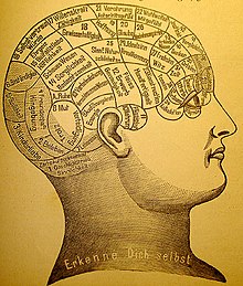
The German anatomist Franz Joseph Gall (1758-1828) found the idea of a functional structure of the brain, as suggested by the Würzburg physician and scholastic Berthold Blumentrost in an Avicenna - Aristotle commentary in the 14th century (1347), brain-topographical in the sense of a Localization theory or localization theory mapped out was the first to be generally hearing. He was not very interested in pathological cases and argued mainly with the anatomical observation that different areas of the cerebrum are connected to differently specialized structures in the brain stem . Gall's lectures were primarily aimed at the interested public; In 1801 he was prohibited from doing this by an imperial edict. It was not until 1809 that he attempted to obtain scientific recognition by submitting it to the French Academy of Sciences . Gall's thesis, in particular, that individual talents and character traits can be traced back to a particularly intense expression of the relevant brain region and that this in turn finds expression in the shape of the skull bone became popular . This cranial science (phrenology) initially developed as a separate branch of science, independent of established brain research, with numerous societies and journals. Over time, however, it turned out that their theories did not match the observations of brain injuries, and their advocates had to neglect more and more cases of healthy people whose abilities did not match the shape of their head. By the middle of the 19th century, phrenology had completely lost its influence and with it not only Gall, but also his completely correct idea of the functional structure was discredited.
19th century
Broad acceptance of the functional localization
After Gall's phrenological research program had failed, a holistic theory first took hold again in the 19th century, the leading proponent of which was Pierre Flourens (1794–1867). She assumed that all sensory impressions and abilities are distributed over the entire brain. With the Parisian doctors Jean-Baptiste Bouillaud (1796–1881) and Simon Alexandre Ernest Aubertin (1825–1893) there were soon voices again that suspected the presence of language in the frontal lobe, but the scientific community still met them with skepticism: There have been too many known cases of frontal lobe injuries that have retained speech.
Paul Broca (1824–1880) made localization theory heard again when he carried out an autopsy on a patient in 1861 ("Monsieur Tan"), who could still understand the language but could no longer express himself linguistically. Broca found a clearly delineated damage in an area of the left frontal lobe, which is still called Broca's area today , and was thus able to specify the location of the language ability more precisely than before. In 1865 it was realized that the left hemisphere plays a special role in speech. Marc Dax (1771-1837) had already expressed this assumption in 1836 and his son Gustave Dax had referred to this discovery in a manuscript that had been received by the Paris Académie de Médecine a few days before Broca went public. Whether Broca was aware of Dax's research when he wrote his own article is considered uncertain. Since abilities returned over time in some patients after brain damage, Broca already postulated mechanisms of cortical plasticity , due to which brain areas can take on tasks that were originally foreign to them, and recommended supportive speech therapy for stroke patients .
After the idea of functional localization was taken seriously again, further findings soon followed: The Englishman John Hughlings Jackson (1835–1911) pointed out the role of the right hemisphere, for example, in spatial orientation and in recognizing people. In 1874, the German doctor Carl Wernicke (1848–1905) found an area in the left temporal lobe, now known as the “ Wernicke Center ”, which is responsible for understanding speech and, if it fails, the ability to speak remains, but no meaningful sentences can be formed . In order to expand the localization theory under controlled conditions, the Germans Gustav Fritsch (1838–1927) and Eduard Hitzig (1838–1907) studied the motor cortex of dogs by means of electrical stimulation and found out in 1870 that certain brain regions are responsible for controlling certain body parts. The function of other regions, especially the frontal lobe, was less easy to determine with electrical stimulation. In such cases, observations after brain injuries were still used: for example, the case of Phineas Gage , who had survived a serious injury to the frontal lobe in 1848, suggested that this plays a role in long-term behavior planning and impulse control, among other things.
David Ferrier (1843–1928) surgically removed certain brain regions from monkeys and gave localization theory its final breakthrough when he demonstrated at an international congress in 1881 that in this way specific functional failures can be induced in a targeted manner. Due to the research of the surgeon Joseph Lister (1827–1912), who introduced the principles of antiseptic surgery, it became possible to keep animals alive for a long time after a brain operation and to observe their behavior. However, this approach provoked sharp protests from animal rights activists, who enforced legal regulation of scientific animal experiments in Great Britain in 1876 . The brain researchers countered these moral concerns by stating that increasing knowledge about cortical localization made it possible to identify the location of brain tumors based on the observable functional failures and thus to save human lives. The first operation on this basis was carried out in 1879 by the Scottish surgeon William MacEwen (1848-1924).
Brain anatomy and cell theory

See also: neuron theory
In the 19th century, research into brain anatomy also made rapid progress. In 1811 Charles Bell (1774–1842) recognized the functional difference between the nerves emerging from the posterior horn and the anterior horn of the spinal cord (dorsal and ventral spinal cord roots ). While motor signals leave the spinal cord via the anterior horn root, sensory signals enter the posterior horn. This discovery was made practically at the same time by François Magendie (1783–1855) and is still in textbooks today as the Bell-Magendie Law . Benedikt Stilling (1810–1879) systematically examined the anatomy of the spinal cord further by making a series of incisions and following the course of various nerve bundles. With the distinction between the corpus geniculatum mediale and the corpus geniculatum laterale of the thalamus , Friedrich Burdach (1776–1847) laid a further foundation stone in the knowledge of the sensory apparatus in 1822, since these two structures should turn out to be the most important switching stations of the hearing and visual senses.
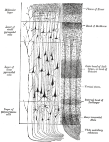
Jules Gabriel François Baillarger (1809–1890) first described in 1840 the 6-layer structure of the gray matter of the cerebral cortex, which is still valid today, and identified a horizontal network of myelinated nerve fibers at the level of layer 4 (Baillarger stripe). The gray matter was thus functionally differentiated and Baillarger then made further considerations about the connection between gray and white matter in the cerebral cortex. Another cornerstone of research is the terminology of cerebral lobes and convolutions proposed by Alexander Ecker (1816–1887) in 1869 and which is still valid today.
More important than the discoveries of macroscopic structures, however, were the discoveries on a small scale. Gabriel Gustav Valentin (1810–1883) identified the nucleus and nucleus of nerve cells for the first time in 1836 . This discovery laid the foundation for the theory published in 1839 by Theodor Schwann (1810–1882) and Matthias Schleiden (1804–1881) that the central nervous system is made up of individual cells. Schwann also identified the cells that form the myelin sheaths around the nerves in the peripheral nervous system (Schwann cells) . Also in the 1830s, Robert Remak (1815–1865) described the different fiber types of myelinated and unmyelinated nerve fibers and suggested that nerve fibers arise from nerve cell bodies. Jan Evangelista Purkinje (1787–1869) also described large neurons in the cerebellum (the so-called Purkinje cells ) in the same years . Purkinje also documented the cellular nature of the tissue layer.
Nevertheless, the cell theory was initially unable to generally prevail against the view that the brain is a continuous ( anastomosic ) mass of tissue, a syncytium . Only an increasing quality of microscopes and better staining methods made it possible to finally clarify this question. The first advances in the preparation of brain tissue for microscopic studies were made by Johann Christian Reil (1759–1813), who used alcohol, and Adolph Hannover (1818–1894), who used chromic acid to harden brain tissue and thus cut thinner sections for microscopy. Alfonso Giacomo Gaspare Corti (1822–1876) initially introduced carmine as a coloring agent, and in 1873 Camillo Golgi (1843–1926) succeeded in coloring with silver nitrate . However, the results were still not subtle enough to dissuade Golgi from his belief that the cells are firmly connected like a pipe system.
Soon, however, there were increasing numbers of indications that spoke in favor of a clear demarcation of the cells: Otto Deiters (1834–1863) described the processes of nerve cells, the dendrites and axons , which were only introduced in 1890 by Wilhelm His (1831–1904) in a posthumously published paper in 1865. and in 1896 by Albrecht von Kölliker (1817–1905) received their current names. The Swiss His and Auguste Forel (1848–1931) argued in their writings in 1886 and 1887, independently of one another, that nerve cells do not fuse with one another. This position was helped by the exceptionally clear preparations that Santiago Ramón y Cajal (1852–1934) made after he had perfected the Golgis staining method, and which showed no firm connection between the cells. Since, as a Spaniard, he was initially an outsider in the scientific community, Heinrich Wilhelm Waldeyer (1836–1921) worked alongside Kölliker for the recognition of his findings and in 1891 established the term “ neuron ”. In 1906 Ramón y Cajal and Golgi were jointly awarded the Nobel Prize for Physiology or Medicine , which was newly established in 1901 . When Golgi came back to the syncytium theory as part of the award and also questioned the functional localization, he was already taking an outsider position.
Ramón y Cajal had already recognized in 1891 that nerves only transmit impulses in one direction and postulated a gap between two nerve cells - which was still invisible at the time - which Charles Scott Sherrington (1857–1952) named " Synapse " in 1897 . In 1891, Sherrington discovered that the patellar tendon reflex is the result of a combination of excitation of an agonist and inhibition of an antagonist . He was able to show that even in animals in which the connection between the brain stem and the cerebrum has been severed, very complex reflexes can still be evoked. His detailed investigations into the neural basis of the reflex refuted the previously common notion that the spinal cord had its own soul. Between 1893 and 1909, Sherrington developed the principles of reciprocal innervation further. He also studied the skin areas that are supplied by individual sensory nerves in monkeys, and published from 1902 together with Albert Grünbaum (1869-1921, from 1915 Albert Leyton) several papers on the precise subdivision of the motor cortex. Sherrington was awarded the Nobel Prize in 1932; since he not only remained scientifically productive for a long time, but also produced a large number of high-profile students, including many others Wilder Penfield (1891-1976), John Carew Eccles (1903-1997) and Ragnar Granit (1900-1991), he had significant influence on brain research well into the 20th century.
Basics of neurology, anesthesia and psychophysics

The French doctor Jean-Martin Charcot (1825–1893), as head of the Paris Hôpital Salpêtrière, laid the foundations of modern neurology: when classifying his patients, he was the first to consistently differentiate between clinically observable symptoms and underlying diseases, the cause of which he sought to clarify by means of autopsies. Together with Edmé Félix Alfred Vulpian (1826-1887) he researched multiple sclerosis : In 1863 he described the typical focal lesions in the central nervous system and differentiated the disease from Parkinson's disease , which was first described by James Parkinson (1755-1824) in 1817 and has been mostly since then had been equated with multiple sclerosis. Charcot also examined amyotrophic lateral sclerosis , led Georges Gilles de la Tourette (1857–1904) in his research into Tourette's syndrome and differentiated epilepsy from hysteria . Only the hysteria research that Charcot carried out towards the end of his scientific career has hardly survived from today's point of view: Charcot deviated from his previously strictly scientific methods and was probably also the victim of his employees, who rewarded patients for displaying certain behaviors .
Great progress was also made in anesthesia in the 19th century: Friedrich Sertürner isolated morphine as a pain reliever in 1803 . Crawford W. Long first used ether as a narcotic in 1842 , Horace Wells (1815–1848) introduced nitrous oxide in 1844, and in 1847 James Young Simpson (1811–1870) successfully used chloroform .
Finally, Ernst Heinrich Weber (1795–1878) and Gustav Theodor Fechner (1801–1887) laid the foundations of psychophysics , the systematic and empirical research into stimulus-experience relationships. Without her groundbreaking research, studying the interaction between objectively measurable physical processes and stimulus strengths and subjective mental experience (external psychophysics) would be impossible. Since the strength of the underlying neuronal processes is mostly more similar to the subjective sensation than the physical stimulus, knowledge of these relationships is crucial for finding and investigating neural correlates of perception (physiology and internal psychophysics).
20th century
Mapping of the cerebral cortex and advances in functional localization
At the turn of the century, Oskar Vogt founded his "Neurological Central Station", initially operated privately in a tenement house, in Berlin , which in 1902 became part of the university and in 1915 became the Kaiser Wilhelm Institute for Brain Research. Together with his wife, the French neurologist Cécile Vogt (née Mugnier), Oskar Vogt had set himself the goal of finding connections between psychological phenomena and anatomical structures of the brain. Part of this project was the work of Korbinian Brodmann , who, as an employee of the institute, published a brain atlas in 1909 in which he divided the cerebral cortex into 52 areas. The basis for the classification were histological examinations in which he stained brain sections with a method developed by Franz Nissl and found differences in the shape and layer thickness of the cells under the microscope . Meanwhile, the Vogts concentrated on the nerve connections between individual areas. Their working hypothesis was that differences in cell architecture indicate brain centers with different functions. In animal experiments, however, they did not succeed in assigning specific functions to the areas Brodmann found. In further research it turned out that such an assignment is actually only possible to a very limited extent; Nevertheless, the Brodmann areas were further refined and they are still used today.
Kurt Goldstein criticized the rigid topographical division of the brain into functional centers (1934).
In 1949 the Nobel Prize was awarded to two researchers who had succeeded in localizing certain functions in the brain. The Swiss physiologist Walter Rudolf Hess had observed the effects of targeted electrical stimulation in the diencephalon of experimental animals and based on these experiments created a detailed functional map of the diencephalon. The second winner was the Portuguese neurologist António Caetano de Abreu Freire Egas Moniz , who introduced leukotomy (also known as “lobotomy”) to treat psychiatric diseases. This surgical procedure, in which parts of the patient's brain tissue were severed, founded psychosurgery . Due to the drastic side effects, however, after the emergence of neuroleptics, it was soon rarely used and the award of the Nobel Prize is now considered a controversial decision.
Another milestone in modern neuroscience was published by Wilder Penfield and Theodore Rasmussen at the beginning of the second half of the 20th century . Using electrical stimulation in the open brain of awake patients, Penfield and Rasmussen tried to locate epileptic centers, but instead triggered complex sensory impressions or spontaneous movements in their patients. Her systematic research into the relationship between the stimulation sites and the movements and sensory impressions revealed a distorted image of the surface of the body, the homunculus, and thus a fundamental principle of brain organization, the somatotopia .
The American neurobiologist Roger Sperry (1913–1994) exchanged certain nerves in adult test animals in the early 1940s and showed that the animals did not get used to this change even after a long time and despite training. These experiments showed how important it was not to randomly connect severed nerves of the motor system to muscles during surgical interventions. After Sperry had read that injured optic nerves regenerate on their own in newts , he removed the eyes of test animals and turned them back in again, turned by 180 °: The nerves nevertheless grew back to their original endpoints, so that the animals' field of vision changed reversed. To explain this, Sperry favored the theory of chemical growth factors that direct nerves to their designated endpoints as they develop. The experimental evidence for such a nerve growth factor was provided in the 1950s by Rita Levi-Montalcini (1909–2012), who was the fourth woman to receive the Nobel Prize in Medicine or Physiology in 1986.
From 1953 Sperry devoted himself to the functional specialization of the two cerebral hemispheres. First he experimented with cats and monkeys, which he cut through the bar that connects the two halves of the brain. As his test animals were recovering from the operation, neurosurgeons Philip Vogel and Joseph Bogen dared to perform a similar operation in 1961 on a war veteran who was suffering from severe and uncontrollable epilepsy. After this treatment method was successful, it was also used in other patients and Sperry extended his experiments to these so-called split-brain patients . The functional division of the two halves of the brain had been speculated since the 19th century. For the first time, Sperry received experimentally verified results that refuted many of the existing convictions: While the right hemisphere was previously generally inferior and incapable of consciousness, Sperry was able to show that it was capable of independent performance and was superior to the left half in certain tasks, for example, when recording space or recognizing patterns and voices. In 1981 he received half of the Nobel Prize for Medicine for his findings.
The second half of the Nobel Prize was shared that year by Torsten N. Wiesel and David H. Hubel , who had contributed to the elucidation of the functioning of the visual cortex with single cell recordings .
One of the most sensational discoveries was made in 1996 by Giacomo Rizzolatti , Leonardo Fogassi and Vittorio Gallese (* 1959), who found cells in the premotor cortex of macaques that became active both when the monkey performed a certain action and when the monkey performed a certain action watched the experimenter perform the same action. With this ability to mirror the actions of other individuals , these so-called mirror neurons have become the basis of a whole series of theories, which primarily seek to explain human cultural achievements (language, ethics).
Coding and transmission of information in the nerves
Although it had been known since the 18th century that electricity played an essential role in conveying information through the human nervous system, the details could not be explored until the 20th century due to a lack of measurement technology. Using a capillary electrometer , Francis Gotch (1853–1913) succeeded in 1899 in observing the refractory period of nerve cells. 1905 showed Keith Lucas (1879-1916), the " all-or-none law " in the activation of muscle cells after, in 1913 his pupil was Edgar Douglas Adrian transfer this knowledge to the activation of nerve, a closer examination of the course of an action potential was initially not possible with the instruments of that time.
The situation changed when the doctor Alexander Forbes (1882–1965) got to know the new tube amplifiers during his military service in World War I and demonstrated after the war that they enabled a substantial and low-distortion amplification of nerve impulses. Herbert Spencer Gasser (1888–1963) constructed a multi-stage amplifier with their help and combined it with a Braun tube . With the oscilloscope created in this way , he and his teacher Joseph Erlanger (1874–1965) examined the temporal course of action potentials for the first time in 1922 and pointed out the different conduction speeds of different nerve cells. In 1944, her research was awarded the Nobel Prize.
Adrian took over the new technology. He succeeded in examining individual cells and, in this way, in 1925, experimentally confirming Sherrington's theory that muscles are deeply sensitive , that is, they transmit information about their tension to the central nervous system via muscle spindles . Together with his student Yngve Zotterman (1898–1982) he found out in 1926 that the intensity of a stimulus is mediated by the frequency of the action potential and that the perceived strength of a persistent stimulus decreases over time ( sensory adaptation ). Even after he and Sherrington had received the Nobel Prize in 1932, Adrian remained scientifically active and in 1943 mapped not only the cerebellar cortex , but also the somatosensory cortex , pointing out that the neural representation of a body part was more of its importance for the survival of the entire organism than corresponds to its size.
For a long time it was debated how nerve impulses spread from one neuron to the next. As early as 1877, Ramón y Cajal had raised the possibility that this stimulus transmission could take place chemically, but did not elaborate on this hypothesis. George Oliver and Edward Albert Sharpey-Schafer (1850–1935) found the first experimental evidence for this when they noticed in 1894 that the injection of an extract of the adrenal gland had an effect on the sympathetic system similar to that of electrical stimulation. In 1905, Thomas Renton Elliott (1877–1961) determined that this effect was specific for the sympathetic nervous system, and concluded from this that the nerve itself could release a substance now called adrenaline . In 1907, Walter Dixon (1871–1931) found a substance similar to muscarine that seemed to play an analogous role for the parasympathetic nervous system. As early as 1906, Reid Hunt (1870–1948) had pointed out that acetylcholine lowered blood pressure significantly, but although he presented this finding at the same congress as Dixon, the two initially made no connection between their results.
The credit for drawing the correct conclusions from the experiments and establishing the view that the transmission of stimuli via the synaptic gap occurs through neurotransmitters goes to Henry Dale (1875–1968) and Otto Loewi (1873–1961), who won the Nobel Prize for this in 1936 received. In 1921, Loewi had obtained a liquid from the excited vagus nerve of a frog that dampened the heartbeat of other frogs and thus provided a clear indication of chemical transmission. Dale showed that the effect of acetylcholine came very close to a natural excitation of the parasympathetic nervous system, and together with Harold Dudley (1887-1935) in 1929 , he demonstrated this substance in the spleen of horses. In 1936 he showed together with Wilhelm Feldberg (1900–1993) that muscles are also activated by neurotransmitters. In 1946 , Ulf von Euler (1905–1983) identified noradrenaline as a messenger substance of the sympathetic nervous system . Together with his colleagues Julius Axelrod (1912–2004) and Bernard Katz (1911–2003), he received the Nobel Prize in 1970 for this.
The Australian John Carew Eccles (1903–1997) was finally able to discover that it is a property of the synapse whether it has a stimulating (excitatory) or inhibitory effect on the downstream cell. He shared it in 1963 the Nobel Prize with two researchers, who solved the exact mechanisms of the electrical stimulus transmission in the nervous system: The British Alan Lloyd Hodgkin (1914-1998) and Andrew Huxley (1917-2012) was in experiments with Riesenaxonen of squid already noticed in 1939 that the membrane potential of a nerve cell in the course of an action potential not only compensates, but reversed: When they their research after the Second world war continued, they could show that this effect on a voltage-dependent permeability of ion channels for sodium - and potassium - ion is based. From this insight they developed the Hodgkin-Huxley model , which enables a realistic simulation of action potentials on the computer.
However, it was only possible to study individual ion channels using the patch-clamp technique developed by the Germans Erwin Neher (* 1944) and Bert Sakmann (* 1942) in 1976. With it they gained knowledge about the behavior of individual membrane proteins , for which they received the Nobel Prize in 1991. In 2000 was followed by a Nobel Prize for three researchers who had contributed to a better understanding of the chemical processes at synapses: The Swede Arvid Carlsson (* 1923), the function of the substance had dopamine as a neurotransmitter and its role in the pathogenesis of Parkinson's disease discovered , while the American Paul Greengard (* 1925) researched the exact sequence of reactions that make up the effect of dopamine.
Numerous theories have also been proposed as to how memory might be encoded in the brain. A series of experiments by the American researcher Georges Ungar seemed to show that fear of the dark is caused by the molecule scotophobin . This theory turned out to be untenable. Eric Kandel (* 1929), also an American, described the mechanisms of synaptic plasticity and their importance for learning and memory processes: Memory is stored in the structure and strength of the connections between nerve cells .
Brain research and ideology
In the first half of the 20th century, brain research also became a political issue. On the one hand, ideas about the organization of the brain often followed the respectively favored social order: Paul Flechsig imagined the cerebrum divided into three strict hierarchy levels based on the model of the monarchy , while Theodor Meynert and Oskar and Cécile Vogt adopted a more republican model, based on the the function of the brain is based on an equal interaction of the individual centers. On the other hand, brain research became a means of political propaganda: In 1925, Oskar Vogt was invited by the Soviet Union to set up a state institute for brain research in Moscow and to dissect the brain of the recently deceased Lenin there. When he summarized his results in a lecture in 1929, he came to the conclusion that the "brain anatomical findings" identify Lenin as an "association athlete" - a conclusion which is daring by today's standards and which has earned him ridicule to this day.
The collaboration of some neuroscientists with those in power during the Nazi era in Germany had far more inhuman consequences . As early as 1920, the psychiatrist Alfred Hoche, together with the criminal lawyer Karl Binding, coined the term “life unworthy of life” and publicly pleaded for the killing of patients who were incurably mentally ill. In his successor, a number of German doctors and neuroscientists helped to (pseudo-) scientifically legitimize and carry out the so-called Action T4 of the National Socialists, under the euphemistic name of " euthanasia ", systematically disabled and psychiatric patients.
Oskar Vogt was withdrawn from the management of the Kaiser Wilhelm Institute in 1935 because he was impartial towards Jews, employed foreigners and, according to the Gestapo, had not acted adequately against “communist propaganda” within his institute. After a transitional period, during which he acted as acting director, Hugo Spatz took over the institute in 1937 in close collaboration with Julius Hallervorden . The two were officially informed about the T4 campaign and received a large number of brains for their research. Hallervorden was present in at least one case when children were murdered in Görden , whose brains were then dissected in his department. Both scientists held leading positions at the Max Planck Institute for Brain Research , the successor organization to the Kaiser Wilhelm Institute, until well into the post-war period , and in some cases continued to research the materials “gained” during the war. An active processing of this dark chapter of brain research in Germany did not begin until the turn of the millennium. The memorial at the Max Delbrück Center in Berlin, which was inaugurated in 2000, and the Prinzhorn Collection established in 2001 at the University of Heidelberg, bear witness to this .
Non-invasive methods enable novel insights
At the beginning of the 20th century, brain researchers were essentially limited to dissecting the brains of the deceased, examining patients with brain damage, or experimenting on the exposed brains of laboratory animals. It was only in the second half of the century that methods emerged that made it possible to conduct examinations on living, healthy brain without having to undertake an intervention. It was these new methods that provided the key to much of the knowledge about the brain that is taken for granted today. The first of these key methods was electroencephalography (EEG), which allows voltage fluctuations on the head surface to be recorded. These in turn allow conclusions to be drawn about the electrical activity of the brain.
As early as 1875, Richard Caton had described a "continuous spontaneous electrical activity" of the brain surface, which he, however, still derived from the exposed brains of rabbits and monkeys. The first recorded EEG was published in 1912 by Vladimir Vladimirovich Pravdich-Neminsky , who still referred to it as "Electrocerebrogram". The name of Hans Bergers , who was the first to record a human EEG in 1924 and published his results in 1929, remained inextricably linked to the procedure . In the years that followed, numerous researchers developed the process further, but it was only used routinely from the 1950s. An important milestone for the comparability of the results achieved was the standardization of the 10-20 system for arranging the EEG electrodes, which was carried out under the direction of Herbert Jasper .
The imaging process made it possible to produce sectional images of the living brain. Due to its suitability for visualizing soft tissue and the low exposure of the test subject, magnetic resonance tomography (MRT) developed in the 1970s , for which Paul Christian Lauterbur and Peter Mansfield received the Nobel Prize in 2003, is in the foreground . Functional magnetic resonance tomography (fMRI), with which the activation of brain centers can be made visible indirectly, has developed into a central method for brain research . As early as 1890, C. S. Roy and C. S. Sherrington had found out that neuronal activity goes hand in hand with a locally increased blood flow to the corresponding brain tissue (so-called " neurovascular coupling "). In 1990, Seiji Ogawa, together with Tso-Ming Lee and other colleagues at Bell Laboratories , made use of the difference in the magnetic properties of oxygen-rich and oxygen-poor blood (" BOLD contrast ") , which has been known since 1935, in order to achieve this increase in the Make MRI images visible.
The diffusion behavior can also be measured by magnetic resonance (in medicine for the first time in 1985 by Denis LeBihan ) and enables conclusions to be drawn about the position and alignment of nerve bundles in the brain. The most common model for this to date, diffusion tensor imaging , was introduced by Peter Basser in 1994 . In practical research, however, this method has so far played a subordinate role compared to fMRI.
Measurements using MRI or EEG only enable correlations to be established between cognitive functions and certain brain activations. In order to be able to research causal relationships between changes in the state of the brain and brain functions, it is necessary, however, to manipulate the brain in a targeted manner and to observe the consequences. A non-invasive way of doing this is by stimulating it with electrical currents that can be induced by strong magnetic fields . As early as 1894 Jacques-Arsène d'Arsonval reported on experiments in which he completely surrounded the head of a test person with a coil through which a strong alternating current flowed and thus triggered, among other things, light sensations, so-called phosphenes . Even if it is controversial today whether d'Arsonval actually stimulated the brain or only the optic nerve and retina of his test subjects, his experiments represent the first attempt to influence the brain using external magnetic fields. In its current form, transcranial magnetic stimulation (TMS) was first described in 1985 by Anthony Barker . He stimulated limited regions of the cortex with much smaller magnetic coils . As a rule, this leads to a temporary disruption of the affected brain area and is associated with functional impairments of the test subject, from the nature of which it is possible to infer the function of the brain area.
Stand at the beginning of the 21st century
In the still young 21st century, neuroscience is developing primarily methodologically. Research into intelligent contrast agents for functional magnetic resonance imaging is being advanced in order to make the concentration (and its change) of any substances in the brain measurable. In principle, these substances should indicate the presence of a certain substance through a change in their magnetic properties that can be measured in the magnetic resonance tomograph. This means that the release of neurotransmitters or the action-potential-coupled influx of calcium into nerve cells in the active, living brain could be followed practically in real time.
Another research direction is the functional study of the neocortex at the cell and synapse level. For this so-called blue brain project, one of the 100 fastest computers in the world, a blue gene supercomputer with 360 teraflops , has been purchased at the Brain Mind Institute in Lausanne , in order to summarize the knowledge gained in a gigantic computer model. Through the exact and systematic study of a single so-called cortical column of 2 mm high and a diameter of 0.5 mm and its 10,000 nerve cells and approximately 10 8 synapses, one hopes the function of the various transmitter, receptor, synapses and cell types in the microcircuits to get on the track of the cortex before the model is extended to the entire cortex (with its approximately 1 million columns). This line of research also benefits from the development of new microscopy techniques that allow real 3D images of entire brains.
A still inconsistent and controversial area within brain research is the search for a neural correlate of consciousness . Although the physicist and biochemist Francis Crick claimed in 1990 that this question could already be dealt with in a meaningful way, and he and Christof Koch presented a corresponding theory, there is still no generally accepted research program to investigate this question (as of 2008). Some philosophers, such as Thomas Metzinger , work on issues that they consider to be a necessary prerequisite for scientific research in this area.
Nevertheless, at the beginning of the 21st century, this topic is particularly present in the press and in popular scientific publications and is used, among other things, as an opportunity to re-pose the question of the compatibility of free will and determinism . In the public debate, some neuroscientists, including Wolf Singer and Gerhard Roth , suggest that future research results will change our image of man and our legal system. Other authors, including Julian Nida-Rümelin , Jürgen Habermas and Michael Pauen , on the other hand, consider it unlikely, based on historical and systematic considerations, that brain research in these areas will cause a fundamental change.
literature
General and historical accounts
- Olaf Breidbach : The materialization of the ego. On the history of brain research in the 19th and 20th centuries. , Suhrkamp, Frankfurt 1997.
- Olaf Breidbach: brain, brain research. In: Werner E. Gerabek , Bernhard D. Haage, Gundolf Keil , Wolfgang Wegner (eds.): Enzyklopädie Medizingeschichte . De Gruyter, Berlin 2005, ISBN 3-11-015714-4 , p. 600 f.
- Peter Düweke: A short history of brain research. From Descartes to Eccles . Becksche series, 2001, ISBN 3-406-45945-5 .
- Matthias Eckoldt : A brief history of the brain and spirit: how do we know how we feel and think . Munich: Pantheon, 2016. ISBN 3570552772 .
- Stanley Finger: Minds behind the brain. A history of the pioneers and their discoveries . Oxford University Press, 2000, ISBN 0-19-518182-4 .
- Michael Hagner : Homo cerebralis. The change from the soul organ to the brain. Insel, Frankfurt 2000, ISBN 3-458-34364-4 .
- Michael Hagner: Ingenious brains. On the history of elite brain research . Munich 2007 (1st edition 2004).
- Michael Hagner: The spirit at work. Historical studies on brain research. Wallstein, Göttingen 2006, ISBN 3-8353-0064-4 .
- ER Kandel , LR Squire: Neuroscience: Breaking Down Scientific Barriers to the Study of Brain and Mind , In: Science , 290: 1113-1120, 2000.
- Erhard Oeser: History of brain research ; Scientific Book Society, Darmstadt 2002.
- Walther Sudhoff: The study of the brain ventricles. In: Sudhoff's archive. Volume 7, 1913, pp. 149-205.
Secondary texts
- Eric R. Kandel, James H. Schwartz, Thomas M. Jessel (Eds.): Principles of Neural Science . McGraw-Hill, New York 2000, 4th Edition, ISBN 0-8385-7701-6 .
- Larry R. Squire, Darwin Berg, Floyd Bloom, Sascha du Lac, Anirvan Ghosh (Eds.): Fundamental Neuroscience . Academic Press; 3rd edition, 2008, ISBN 0-12-374019-3 .
Web links
Individual evidence
- ^ JH Breasted: The Edwin Smith surgical papyrus. Univ. Chicago Press, Chicago 1980, 2 vols. Pp. Xvi, 6, 480-485, 487-489, 446-448, 451-454, 466; 2: S. XVII, XVII (A)
- ^ Stanley Finger: Minds behind the brain. P. 18.
- ↑ Lloyd, 1975 .: Alcmeon and the early history of dissection , Sudhoffs Archiv, 59: 113-47
- ^ Corpus Hippocraticum: De morbo sacro .
- ↑ H. Diels, W. Kranz: The fragments of the pre-Socratic. 6th ed., Volume 1, pp. 210-216. Weidmann, Dublin, Ireland 1952.
- ^ Bernhard D. Haage: Ventrikellehre. In: Werner E. Gerabek , Bernhard D. Haage, Gundolf Keil , Wolfgang Wegner (eds.): Enzyklopädie Medizingeschichte. De Gruyter, Berlin / New York 2005, ISBN 3-11-015714-4 , p. 1439.
- ↑ Olaf Breidbach : Hirn, brain research. In: Werner E. Gerabek , Bernhard D. Haage, Gundolf Keil , Wolfgang Wegner (eds.): Enzyklopädie Medizingeschichte. De Gruyter, Berlin / New York 2005, ISBN 3-11-015714-4 , p. 600 f .; here: p. 600.
- ↑ CM Goss: On anatomy of nerves by Galen of Pergamon. On J Anat. March 1966; 118 (2): pp. 327-335.
- ↑ See also Jost Bendedum : The olfactory organ in ancient and medieval brain research and the reception by S. Th. Soemmerring. In: Gunter Mann, Franz Dumont (Ed.): Brain - Nerves - Soul. Anatomy and physiology in the context of S. Th. Soemmerring. Stuttgart 1988 (= Soemmerring-Forschungen. Volume 3), pp. 11-54.
- ↑ Archived copy ( Memento of the original from September 28, 2007 in the Internet Archive ) Info: The archive link was inserted automatically and has not yet been checked. Please check the original and archive link according to the instructions and then remove this notice.
- ^ L. Richter-Bernburg: Abu Bakr Muhammad al-Razi's (Rhazes) medical works. Med Secoli. 6 (2): pp. 377-392, 1994
- ↑ FS Haddad Surgical firsts in Arabic medical literature. Stud Hist Med Sci. 10-11: pp. 95-103. 1986-1987
- ↑ AS Sarrafzadeh, N. Sarafian, A. von Gladiss, AW lower Berg, WR Lanksch: Avicenna (Avicenna). Historical note. Neurosurg Focus. 11 (2): E5. 2001
- ↑ J. Hirschberg: History of Ophthalmology , Vol. 3, Leipzig 1915.
- ^ E. Savage-Smith: The practice of surgery in Islamic lands: myth and reality. Soc Hist Med. 13 (2): pp. 307-321. 2000
- ^ Jean C. Tamraz and Youssef G. Comair: Atlas of Regional Anatomy of the Brain Using MRI . Springer, 2006, page 1.
- ↑ Leonardo da Vinci on the Brain and Eye. P. 103 , accessed on November 23, 2017 (English).
- ^ Jean C. Tamraz and Youssef G. Comair: Atlas of Regional Anatomy of the Brain Using MRI . Springer, 2006, page 3.
- ^ M. Driver: Bartholomeo Eustachio-the third man: Eustachius published by Albinus. ANZ J Surg. 73 (7): pp. 523-528; 2003
- ↑ PC Kothary, SP Kothary: Gabriele Fallopio. Int Surg. 60 (2): pp. 80-81. 1975
- ↑ Dall'Osso E. A contribution to the scientific thought of Giulio Cesare Aranzio: his surgical work. 1956 61 (6): pp. 754-767 (Italian).
- ^ Stanley Finger: Minds behind the brain. Page 75.
- ^ Antonio R. Damasio. Descartes' error. Feeling, Thinking and the human brain. List, 1997.
- ^ Stanley Finger: Minds behind the brain. Page 102.
- ^ Stanley Finger: Minds behind the brain. Page 121.
- ^ Gundolf Keil: Berthold Blumentrost. In: Werner E. Gerabek, Bernhard D. Haage, Gundolf Keil, Wolfgang Wegner (eds.): Enzyklopädie Medizingeschichte . De Gruyter, Berlin 2005, ISBN 3-11-015714-4 , p. 189 f.
- ↑ Rüdiger Krist: Berthold Blumentrosts 'Quaestiones disputatae circa tractatum Avicennae de generatione embryonis et librum meteorum Aristotelis'. A contribution to the scientific history of medieval Würzburg. Part I: Text. (Medical dissertation Würzburg) Wellm, Pattensen 1987, now published by Königshausen & Neumann, Würzburg (= Würzburg medical historical research. Volume 43).
- ↑ Avicenna already warned against placing cupping heads on the occiput so that the memory would not be damaged. See Gotthard Strohmaier : Avicenna. Beck, Munich 1999, ISBN 3-406-41946-1 , p. 75.
- ↑ The five senses ( auditus , visus , olfactus , gustus and tactus ) as well as sensus communis , fantasya , ymaginacio and cogitacio as well as memoria are differentiated .
- ^ Olaf Breidbach: The materialization of the ego. Page 73.
- ^ Stanley Finger: Minds behind the brain. Page 145.
- ^ Stanley Finger: Minds behind the brain. Page 172.
- ↑ Ramón y Cajal, Nobel Lectures: Physiology or Medicine (1901-1921) (Elsevier, Amsterdam, 1967), pp. 220-253.
- ^ MR Bennett and PMS Hacker : Philosophical Foundations of Neuroscience. Blackwell Publishing, 2003, p. 41, ISBN 1-4051-0838-X
- ^ Stanley Finger: Minds behind the brain. Page 195.
- ↑ Wolf Singer : On the way inwards. 50 years of brain research in the Max Planck Society. In: The Observer in the Brain. Essays on Brain Research . Suhrkamp, 2002, p. 12, ISBN 3-518-29171-8
- ^ Herbert Olivecrona: The Nobel Prize in Physiology or Medicine 1949, Presentation Speech. In: Nobel Lectures, Physiology or Medicine 1942–1962. Elsevier Publishing Company, Amsterdam 1964.
- ↑ Wilder Penfield, Theodore Rasmussen: The Cerebral Cortex of Man. A Clinical Study of Localization of Function. The Macmillan Comp., New York 1950.
- ↑ RW Sperry, Proc. Natl. Acad. Sci. USA 50, 703, 1963
- ↑ DH Hubel and TN Wiesel, J. Physiol. 148, 574, 1959
- ↑ Giacomo Rizzolatti et al. (1996): Premotor cortex and the recognition of motor actions. In: Cognitive Brain Research 3, pp. 131-141.
- ^ Ragnar Granit : The Nobel Prize in Physiology or Medicine 1963, Presentation Speech In: Nobel Lectures, Physiology or Medicine 1963–1970 , Elsevier Publishing Company, Amsterdam, 1972
- ^ Louis Neal Irwin (2006) "Scotophobin: Darkness at the Dawn of the Search for Memory Molecules", ISBN 0-7618-3580-6
- ^ Howard Eichenbaum: Memory . In: Scholarpedia . (English, including references)
- ↑ Peter Düweke: Brief history of brain research. Page 124
- ↑ Peter Düweke: Brief history of brain research. Page 116.
- ^ Hans-Walter Schmuhl : Medicine in the Nazi era: brain research and the murder of the sick. In: Deutsches Ärzteblatt 98 (19), page A 1240-1245, May 2001
- ↑ Speech by President Hubert Markl on the unveiling of the memorial in memory of the victims of the National Socialist euthanasia measures ( Memento of June 7, 2007 in the Internet Archive ) Berlin-Buch, October 14, 2000
- ↑ Barbara E. Swartz and Eli S. Goldensohn: Timeline of the history of EEG and associated fields. In: Electroencephalography and clinical Neurophysiology. 106, 1998, pp. 173-176.
- ↑ LA Geddes: d'Arsonval, Physicial and Inventor. In: IEEE Engineering in Medicine and Biology. July / August 1999, pp. 118-122.
- ↑ AT Barker, R. Jalinous, IL Freeston: Non-invasive magnetic stimulation of human motor cortex. The Lancet 1985; 1: pp. 1106-1107.
- ^ Henry Markram (2006) The blue brain project. In: Nat Rev Neurosci ; 7 (2): pp. 153-160. Review.
- ↑ HU Dodt et al. (2007): Ultramicroscopy: three-dimensional visualization of neuronal networks in the whole mouse brain. In: Nature Methods 4, pp. 331-336.
- ↑ Wolf Singer: A New Image of Man? Talks about brain research. Suhrkamp, 4th edition 2003, ISBN 978-3-518-29196-2 .
- ↑ Michael Pauen: What is man? The discovery of the nature of the mind. Deutsche Verlags-Anstalt, 2007, ISBN 978-3-421-04224-8 .
- ↑ Cf. Jens Elberfeld: Review of: Hagner, Michael: The spirit at work. Historical studies on brain research. Göttingen 2006 . In: H-Soz-u-Kult , March 24, 2010.


