neck
The neck , anat. Collum ( lat. Collum "neck") or cervix (lat. Cervix "neck") is that part of the body of humans and animals that connects the head and torso . With its various functions to be fulfilled, it is a complex structure that also represents a bottleneck at risk.
Belonging to the neck is denoted by the adjective cervical , e.g. B. the term cervical syndrome . However, the cervical can also refer to other anatomical structures, see Cervix uteri (cervix).
etymology
The common. Body part designation mhd. , Ahd. Neck , like lat. Collum, belongs to the idg. Root * ku̯el "[to] turn, [to] move around" and therefore actually means "[head] -twist".
anatomy

Limitations
The border of the neck forms
- Top: the connecting line from the lower border of the mandible to the mastoid process and along the superior nuchal line to the external occipital protuberance of the occipital bone.
- downwards: the line connecting the upper edge of the sternum ( manubrium sterni ), over the collarbone to the acromion of the shoulder blade and backwards to the spinous process of the seventh cervical vertebra.
Regions
In humans, the neck is divided into eight regions ( Regiones cervicales ):
- Regio cervicalis posterior , syn.regio nuchae, neck
- Regiones cervicales ventrolaterales
- Regio cervicalis lateralis (lateral neck area) with trigonum omoclaviculare ( syn.Fossa supraclavicularis major )
- Sternocleidomastoid region with the minor supraclavicular fossa
-
Anterior cervical region (anterior neck region)
- Child triangle ( Trigonum submentale )
- Lower jaw triangle ( Trigonum submandibulare )
- Carotid triangle ( Trigonum caroticum )
- Muscle triangle ( Trigonum musculare ).
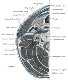
Fascia of the neck

The cervical fascia is divided with its three leaves the neck into a plurality of regions and covering the muscles of the neck. In addition, the various cervical viscera and the ducts of the neck are covered by their own connective tissue structures, the intestinal fascia and the carotid vagina .
The superficial sheet (the lamina superficialis ), as part of the superficial body fascia, envelops the entire front neck, covers the sternocleidomastoid muscle and the parotid gland . Only the platysma and epifascial veins and nerves are still above this fascia. It emerges from the parotid masseterica fascia on the lower jaw , merges downward into the thoracic fascia and towards the neck into the nuchal fascia .
The middle leaf, the lamina pretrachealis, begins around the hyoid bone and extends to the sternum and collarbone. It becomes wider and wider towards the bottom and is particularly firm in the area where it covers the infrahyoid muscles . It is fused with the carotid vagina, the sheath of connective tissue that encases important conduction pathways such as the carotid artery, the internal jugular vein and the vagus nerve .
Under the lamina pretrachealis lies the intestinal fascia, which covers the throat viscera, namely the larynx, pharynx, thyroid, trachea and esophagus.
The deep leaf ( lamina prevertebralis ) of the cervical fascia lies directly in front of the spine and splits downwards so that it envelops the scalene muscles and the prevertebral muscles , the autochthonous neck muscles and the levator scapulae muscle . It extends from the base of the skull to about the third thoracic vertebra, where it merges into the endothoracic fascia . It also covers the sympathetic trunk with the three cervical ganglia, the brachial plexus , the subclavian artery and the phrenic nerve .
Musculature
The muscles of the neck can be divided into different groups. On the one hand, these are the infrahyoid muscles consisting of sternohyoid muscle , sternothyroid muscle , thyrohyoid muscle and omohyoid muscle . These muscles are all important for swallowing. They all shift the hyoid bone down and - apart from the thyrohyoideus muscle - also the larynx and thus also play a role in phonation . The thyrohyoid muscle, on the other hand, moves the larynx upwards, provided the hyoid bone is fixed. The omohyoideus muscle also stretches the pretracheal lamina of the cervical fascia, with which it is fused via its intermediate tendon.
The counterpart to the infrahyoid muscles is formed by the suprahyoid muscles with the digastric muscle , geniohyoid muscle , mylohyoid muscle and the stylohyoid muscle . These muscles lift the hyoid bone when swallowing and also support the opening of the jaw.
The group of the deep neck muscles forms the prevertebral muscles , which results from the musculus longus capitis , the musculus longus colli , the musculus rectus capitis anterior and out of the musculus rectus capitis lateralis composed. You can turn the cervical spine or, if you are acting on both sides, bend it forwards, or you can turn your head to the side or bend it forwards.
In addition, a distinction can be made between the scaleni muscles ("stair muscles"), which consist of the anterior , medius , posterior and, in some people, a scalalenus minimus muscle . These muscles act as auxiliary breathing muscles , or - if the ribs are fixed - can bend the cervical spine to the side or forwards.
The sternocleidomastoid muscle and the superficial platysma cannot be assigned to any group. The neck muscles are also located in the neck, but they are part of the autochthonous back muscles .
Ducts
Arteries
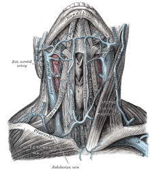
Several large blood vessels run through the neck, some of which are involved in supplying the neck, but some of them without a supply function to the brain. The two vessels of the neck from which all other vessels originate are the common carotid artery and the subclavian artery . On the right these two arteries arise from the brachiocephalic trunk , on the left directly from the aorta . The arteria subclacia does not extend to the neck, but gives off the arteria vertebralis , which runs through holes in the spinous processes of the cervical vertebrae ( foramina transversaria ) up to the skull. It also gives off the thyrocervical trunk , which divides into a series of arteries that essentially supply lateral structures at the base of the neck.
The common carotid artery branches in its further course into the internal carotid artery and the external carotid artery , of which the internal carotid artery does not have a supply function for the neck, but rather leads to the brain. As a rule (50%), the arteria carotis externa, the arteria thyroidea superior , the arteria lingualis and the arteria facialis above the bifurcation emerge individually. From these, the arteria thyriodea superior branches into further arteries, which supply the larynx, the thyroid gland and the musculus sternocleidomastoideus. In addition, a branch of the arteria thyroidea superior - the ramus infrahyoideus - anastomoses with the branch on the opposite side, so that a connection between the two arteries is established. The lingual artery mainly supplies the tongue and the floor of the mouth, but also the base of the tongue and the epiglottis via its rami dorsales linguae .
Further arteries from the external carotid artery are the ascending pharyngeal artery , the posterior auricular artery , the occipital artery , the maxillary artery and the superficial temporal artery , of which only the ascending pharyngeal artery supplies part of the neck via its pharyngeal branch of the larynx.
Veins
The veins in the neck are mostly "thoroughfares" that carry the blood from the head back to the heart. They have no valves, are - as they are above the heart - only slightly filled and are usually not visible when standing. They can only be recognized when lying down in healthy individuals and also when standing in patients with right heart failure . The outflows on both sides of the neck are connected by the arcus venosus jugularis . Care must be taken during tracheostomies because of the risk of bleeding. In addition, the veins are strongly anastomosed to one another so that blood congestion does not occur even if a larger vein is tied.
The largest vein in the neck is the internal jugular vein . It emerges from the cranial cavity through the jugular foramen and thus drains the blood from the brain via the venous outflows of the hard meninges - the sinus durae matris - and via the vena facialis , the vena lingualis , the vena thyroidea superior and the venae thyroideae mediae also blood from the face and thyroid gland.
The external jugular vein drains blood from the superficial area behind the ear. It initially runs over the fascia ( lamina superficialis ), but under the platysma, it then breaks through to open into the subclavian vein .
The anterior jugular vein is variable and begins, when it is pronounced, under the hyoid bone . It runs downwards, drains blood from the anterior, superficial neck region and mostly flows into the external jugular vein.
The subclavian vein finally connects with the internal jugular vein to form the brachiocephalic vein , into which the plexus thyroidus impar and the vena vertebralis also open . The inferior thyroid vein can also flow into it. The thyriodeus impar plexus is a venous plexus that drains blood from the thyroid gland. The connection of the left subclavian vein and internal jugular vein creates the left vein angle , into which the thoracic duct opens. The right and left subclavian veins eventually connect to form the superior vena cava that carries blood to the heart.
There are also a large number of other veins that are too variable, so that they are not mentioned here.
Lymphatics
In general, one can differentiate between superficial and deep lymph nodes and regional and collective lymph nodes. Regional lymph nodes are the first stations in the lymphatic system and receive the lymph from an organ or region. Collective lymph nodes are downstream stations and receive their lymph from regional lymph nodes. Superficial lymph nodes are mostly regional and deep lymph nodes are mostly collective lymph nodes. The lymph nodes flow from the superficial lymph nodes, the nodi occipitales on the occiput, the nodi infraauriculares behind the ear, the nodi parotidei superficialis , the nodi parotidei profundi on the parotid gland, the nodi anteriores superficialis and the nodi laterles superficiales to the deep and collecting nodes the right or left jugular trunk - larger lymph trunks along the internal jugular vein. On the right side the jugular trunk finally opens into the ductus lymphaticus dexter , which ends in the right vein angle, and on the left side the left trunkus jugularis opens into the ductus thoracicus, which ends in the left vein angle.
The deep lymph nodes can be divided into six different regions (according to the American Academy of Otolaryngology ):
- The submetales and submandibular nodes
- The nodi cervicales profundi , each of which can be subdivided into three further groups, namely the upper, middle and lower group
- The Nodi trigoni cervicalis posterioris
- The nodi cervicales anteriores
annoy
The nerves that run in the neck area can be divided into three groups:
- There are the nerves of the cervical spinal cord segments C1 to C4, the anterior branches of which form the cervical plexus and large parts of the brachial plexus . For the posterior branches of the spinal nerves, see the neck , as they innervate its muscles
- In addition, various cranial nerves and nerves run through the neck
- Nerves of the neck part of the sympathetic trunk , which lie in the lamina prevertebralis .
The spinal nerves of the spinal cord segments C1 to C4 give after leaving the spinal cord anterior and posterior branches off, from which the front - after they have given some direct muscular branches - form the cervical plexus. This has both sensory and motor components. The sensitive parts leave the plexus again and pull to the surface at the rear edge of the sternocleidomastoid muscle ( punctum nervosum ). These are the nervus auricularis magnus , the nervus occipitalis minor , the nervus transversus colli and the supraclavicular nerves ( nervus supraclavicularis anterior , intermedius and lateralis ). An exception are the sensitive parts of the phrenic nerve (mostly C4), which is made up of segments C3 to C5. It pulls down to the chest and abdomen and innervates the diaphragm with motor and sensitivity and the peritoneum , pericardium and parietal pleura sensitively.
From the spinal nerves of segments C5 to C8, together with Th1, the brachial plexus arises, which runs through the posterior scalenus gap and over the clavicle. It consists of three branches ( trunci ) that come together and innervate the upper extremity.
The cranial nerves that run in the neck area are the VII ( facial nerve ), IX ( glossopharyngeal nerve ), X ( vagus nerve ), XI ( accessory nerve ) and XII ( hypoglossal nerve ). Only the branches that are important for the structures of the neck are listed here: The facial nerve runs from the rear edge of the lower jaw to the transversus colli nerve and forms a kind of loop there, which was previously called the ansa cervicalis superficialis . The fibers of the facial nerve are of a purely motor nature, while those of the transversus colli nerve are sensory. The VII cranial nerve innervates the platysma in the neck area.
The glossopharyngeal nerve contains mixed fibers, of which the sensory fibers run to the point of division of the common carotid artery, where they innervate the carotid sinus and carotid body with its chemo- and pressoreceptors. In addition, the glossopharyngeal nerve releases sensory and motor fibers in branches ( rami paryngei ) which, together with the pharyngeal rami of the vagus nerve, form the pharyngeal plexus , which supplies almost all the muscles of the pharynx with motor and the entire pharynx. Only the stylopharyngeus muscle is innervated by the motor branches of the glossopharyngeal nerve.
In addition to the innervation of the throat, the vagus nerve is also involved in the innervation of the larynx. For this he gives off two branches: the first branch that Ramus superior laryngeal , supplied the larynx with its Ramus externus the muscle cricothyroideus and his Ramus internus the mucous membrane of the larynx above the glottis Rima. Below the rima glottidis, the innervation of the mucous membrane, as well as that of all internal larynx muscles, belongs to the recurrent laryngeal nerve - the other branch of the vagus nerve that is important for the larynx.
The accessory nerve is purely motoric and partially supplies the sternocleidomastoid and trapezius muscles. The hypoglossal nerve is also motoric and innervates the tongue muscles and the styloglossus muscle .
Further anatomy
Various supply lines such as the esophagus , windpipe and blood vessels run through the neck . The skeleton ( cervical spine ) must produce the greatest possible flexibility for the head. The front part of the neck, which contains the larynx and the pharynx , is called the gargle (from Latin: gurgulio = throat, throat , windpipe).
Idioms

To turn someone 's neck or cut off their neck (literally) means to kill someone as it will disrupt all vital body functions. The same thing happens with executions by means of beheading ( guillotine ) or hanging ("death by hanging "). The idiom is used as an "empty threat" to signal how seriously you mean something.
A cutthroat is someone who financially takes advantage of another, a usurer or an exploiter .
The expression hit someone's throat (or even the throat ) is also a life-threatening attack aimed primarily at interrupting the air supply.
The expression have a frog in the throat has its etymological origin in the frog tumor , medicinally ranula . The swelling of the neck, which is caused by a reddening of the almonds, provides pain when swallowing .
The term having a throat or having a throat describes a state of anger or indignation about a certain condition or the behavior of a person.
Getting a long neck or having a long neck means that someone is stretching or craving something .
The desire to break a neck or a leg originally has nothing to do with the neck, but is a corruption of the Yiddish saying "hatslokhe u brokhe" ("success (luck) and blessing")
The term freedom has its origin in the freedom of the neck, the collum liberum , a neck that has no yoke on itself.
See also
- collar
- a necklace
- Torticollis (torticollis)
- Tooth neck
Individual evidence
- ^ The dictionary of origin (= Der Duden in twelve volumes . Volume 7 ). 5th edition. Dudenverlag, Berlin 2014 ( p. 363 ). See also DWDS ( "neck" ) and Friedrich Kluge : Etymological dictionary of the German language . 7th edition. Trübner, Strasbourg 1910 ( p. 190 ).
- ↑ a b Michael Schünke , Erik Schulte , Udo Schumacher : PROMETHEUS . 4th edition. Head, Neck and Neuroanatomy. Thieme, Stuttgart New York 2015, ISBN 978-3-13-139544-3 , pp. 4 .
- ^ A b Gerhard Aumüller, Gabriela Aust, Andreas Doll, et al .: Anatomie (= dual series ). 2nd Edition. Thieme, Stuttgart 2010, ISBN 978-3-13-136042-7 , p. 804 .
- ↑ Federative Committee on Anatomical Terminology: Terminologia anatomica. Thieme, 1998, ISBN 9783131143617 , p. 3.
- ↑ a b c d e Michael Schünke, Erik Schulte, Udo Schumacher: PROMETHEUS . 4th edition. Head, Neck and Neuroanatomy. Thieme, Stuttgart New York 2015, ISBN 978-3-13-139544-3 , pp. 4 and 5 .
- ↑ a b c d e Gerhard Aumüller, Gabriela Aust, Andreas Doll, et al .: Anatomie (= dual series ). 2nd Edition. Thieme, Stuttgart 2010, ISBN 978-3-13-136042-7 , p. 804 and 805 .
- ↑ a b c d e f Michael Schünke, Erik Schulte, Udo Schumacher: PROMETHEUS . 4th edition. Head, Neck and Neuroanatomy. Thieme, Stuttgart New York 2015, ISBN 978-3-13-139544-3 , pp. 88-93 .
- ↑ a b c d e Gerhard Aumüller, Gabriela Aust, Andreas Doll, et al .: Anatomie (= dual series ). 2nd Edition. Thieme, Stuttgart 2010, ISBN 978-3-13-136042-7 , p. 806-808 .
- ↑ Michael Schünke, Erik Schulte, Udo Schumacher: PROMETHEUS - learning atlas of anatomy . 4th edition. Head, Neck and Neuroanatomy. Thieme, Stuttgart New York 2015, ISBN 978-3-13-139544-3 , pp. 94 .
- ↑ Michael Schünke, Erik Schulte, Udo Schumacher: PROMETHEUS - learning atlas of anatomy . 4th edition. Head, Neck and Neuroanatomy. Thieme, Stuttgart New York 2015, ISBN 978-3-13-139544-3 , pp. 94-99 .
- ^ Gerhard Aumüller, Gabriela Aust, Andreas Doll, et al .: Anatomie (= dual series ). 2nd Edition. Thieme, Stuttgart 2010, ISBN 978-3-13-136042-7 , p. 809 .
- ↑ Michael Schünke, Erik Schulte, Udo Schumacher: PROMETHEUS - learning atlas of anatomy . 4th edition. Head, Neck and Neuroanatomy. Thieme, Stuttgart New York 2015, ISBN 978-3-13-139544-3 , pp. 94 and 99 .
- ↑ a b c d e Michael Schünke, Erik Schulte, Udo Schumacher: PROMETHEUS - learning atlas of anatomy . 4th edition. Head, Neck and Neuroanatomy. Thieme, Stuttgart New York 2015, ISBN 978-3-13-139544-3 , pp. 108 and 109 .
- ↑ a b Michael Schünke, Erik Schulte, Udo Schumacher: PROMETHEUS - learning atlas of anatomy . 4th edition. Head, Neck and Neuroanatomy. Thieme, Stuttgart New York 2015, ISBN 978-3-13-139544-3 , pp. 110 .
- ↑ a b c Gerhard Aumüller, Gabriela Aust, Andreas Doll, et al .: Anatomie (= dual series ). 2nd Edition. Thieme, Stuttgart 2010, ISBN 978-3-13-136042-7 , p. 813 and 814 .
- ^ Gerhard Aumüller, Gabriela Aust, Andreas Doll, et al .: Anatomie (= dual series ). 2nd Edition. Thieme, Stuttgart 2010, ISBN 978-3-13-136042-7 , p. 418 .
- ^ Gerhard Aumüller, Gabriela Aust, Andreas Doll, et al .: Anatomie (= dual series ). 2nd Edition. Thieme, Stuttgart 2010, ISBN 978-3-13-136042-7 , p. 815 and 816 .
- ↑ a b c Gerhard Aumüller, Gabriela Aust, Andreas Doll, et al .: Anatomie (= dual series ). 2nd Edition. Thieme, Stuttgart 2010, ISBN 978-3-13-136042-7 , p. 816 .


