diaphragm
| Diaphragm ( diaphragm ) |
|---|

|
| origin |
| Lumbar part ( pars lumbalis ): lumbar vertebrae Rib part ( pars costalis ): inside of the seventh to last rib |
| approach |
| Centrum tendineum |
| function |
| Inspiration (inhalation) |
| Innervation |
| Phrenic nerve from the cervical plexus |
| Spinal segments |
| C3-C5 |
The diaphragm or diaphragm (from ancient Greek διάφραγμα diaphragm "partition wall" or "diaphragm") is a muscle - tendon -Platte which in mammals the chest and the abdominal cavity from each other. It has a dome- shaped shape and is the main breathing muscle . The muscle contraction of the diaphragm leads to inhalation ( inspiration ). In humans, it is 3 to 5 mm thick and does 60 to 80% of the muscle work required for inspiration at rest.
Apart from mammals, only crocodiles have a structure comparable to the diaphragm .
Explanations of words
The name "Zwerchfell" is derived from the outdated German word zwerch ("quer"). The component "fur" comes from Germanic * fel for "Skin" ( Indo-European * pel . See Pelle , peel up for "molt") from, have the same meaning in the eardrum , peritoneum or animal fur . Since laughter is a strongly accelerated sequence of breathing movements and the diaphragm is involved in this process, there are a number of related phrases and word combinations in German ("diaphragm attack").
In ancient Greece , the diaphragm was believed to be the seat of the soul , which is why the word phrēn (φρήν) stands for both terms. Hence the word comes in the name of mental illness Schizo phren ie before, although the diaphragm is not involved in this disease.
The medical term diaphragm comes from the late Latin diaphragm "diaphragm", which comes from the ancient Greek διάφραγμα ([ dɪˈapʰraŋma ]), which means "partition, septum" and "diaphragm". The anatomical term comes from Gerard von Cremona . It is used both in anatomy for additional partition walls through which something can pass, such as in the pelvic floor ( diaphragm pelvis and diaphragm urogenitale ) or the diaphragm sellae (between the base of the brain and pituitary gland ) and outside the anatomy (→ diaphragm ).
Anatomical structure
The muscular part of the diaphragm is divided into three parts according to its origin : lumbar, sternum and rib part. All three parts end in a common tendon plate ( centrum tendineum ), which consists of the intertwined tendon fibers . The ratio between the muscular and the sinewy part is variable within the mammals. Dogs and cats have a small, narrow and Y-shaped tendon plate, in other domestic animals and in humans it is V -shaped to heart-shaped and represents the largest portion of the diaphragm.
The lumbar part ( pars lumbalis ) arises on the ventral side ( ventral ) of the lumbar spine . It consists of a right leg ( crus dextrum ) and a left leg ( crus sinistrum ). These "diaphragmatic pillars" represent muscle strands that pull upwards in humans and forward in animals as a result of the horizontal body orientation. The right thigh is stronger and can be divided into two (human: crus mediale and crus laterale ) or three subsections. There are three sinewy arches on the loin part. The quadratus arcade ( ligamentum arcuatum laterale ) and the psoasarcade ( ligamentum arcuatum mediale ) encompass the two parts of the muscle iliopsoas on the abdomen , the aortic arcade ( ligamentum arcuatum medianum ) the aortic slot (see below).
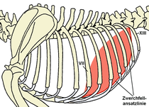
The loin part laterally borders on the rib part ( pars costalis ), which arises on the ribs ( costae ) or on the costal cartilage. The line connecting the individual origins on the ribs ( diaphragm attachment line) runs diagonally and is different in the individual mammal species due to the varying number of ribs. As a guide, it runs on the back ( dorsal ) from the last rib ventrally ( ventrally ) to the seventh rib. The degree of inclination varies according to animal species, i.e. depending on the length of the chest or the number of ribs. In some marine mammals ( whales , manatees ) the diaphragm is almost horizontal. Since the lungs can expand into the space to the side of the apex of the diaphragmatic dome up to this line of attachment ( recessus costodiaphragmaticus ), the line of attachment of the diaphragm also determines the maximum lung percussion field .
The rib part borders on the abdomen on the small sternum part ( pars sternalis ). It arises at the end of the breastbone ( sternum ), at the so-called sword extension ( processus xiphoideus ).
The diaphragm is of a fascia covers and on the thoracic cavity side of the pleura ( diaphragmatic pleura ), on the abdominal cavity side of the peritoneum ( peritoneum coated). The dome shape of the diaphragm results from the negative pressure in the pleural cavity and the effort of the lungs to contract ("retraction force" of the lungs).

1 rib part of the diaphragm, 2 right thighs of the lumbar part of the diaphragm, 3 centrum tendineum of the diaphragm, 4 aorta, 5 thoracic duct, 6 esophagus, 7 right lung (caudal lobe), 8 caudal vein, 9 Phrenic nerve, 10 right lung (accessory lobe), 11 heart in the pericardium, 12 arcus lumbocostalis (Bochdalek gap), 13 last, 13th rib, 14 costal arch, 15 liver, 16 duodenum
There are three larger openings in the diaphragm. The aortic slit ( hiatus aorticus ) is located on the back between the two legs of the loin part. It is arranged at an angle and in humans extends from the first lumbar vertebra to the eleventh thoracic vertebra . The main artery ( aorta ) and a large lymphatic trunk , the thoracic duct, pass through the aortic slit . The esophageal slit ( hiatus oesophageus ) lies between the subsections of the right lumbar leg, in humans at the level of the tenth thoracic vertebra. Through the esophagus slot pull the gullet ( esophagus ) and the two main branches of the vagus nerve ( vagal trunk anterior and posterior , with animals as vagal trunk ventral and dorsal referred). The third larger opening is the vena cava hole ( foramen venae cavae ). It is located in the tendon plate. The inferior vena cava ( inferior vena cava , in animals caudal vena cava ) passes through this hole . In contrast to the two slits in which the structures that pass through are slidably mounted, the vena cava is fused with the diaphragm in a firm ring of connective tissue. This adhesion is responsible for the change in shape of the diaphragm during contraction and prevents the vein from collapsing.
There are also some smaller openings. In the right loin thigh there is an opening for the azygos vein and the major splanchnic nerve . Back side of the Aortenschlitz occurs sympathetic chain ( sympathetic trunk ) through the diaphragm. Muscle- free areas are only closed by loose connective tissue between the muscular parts . The Larrey cleft ( Trigonum sternocostale sinistrum ) and the Morgagni hole ( Trigonum sternocostale dextrum ) lie on the left and right between the rib and sternum. The vena epigastrica superior (in animals, vena epigastrica cranialis ), the terminal branch of the internal thoracic vein ( vena thoracica interna ), runs through it . The Bochdalek gap ( Trigonum lumbocostale , in the veterinary anatomy Arcus lumbocostalis ) lies between the rib and loin parts . This is where the diaphragm is weakest, which is why it is here that collections of pus (abscesses) break through or diaphragmatic ruptures occur. Further weak points are the aortic and esophageal slits, since here too only loose connective tissue stabilizes the opening.
On the chest cavity side, the diaphragm borders on the lungs , the mediastinum and, in some mammals (humans, predators ), also on the pericardium, on the abdominal cavity side above all on the liver . In ruminants , only the right side of the diaphragm has contact with the liver, the liver is displaced to the right by the large forestomach and the left half of the diaphragm is in direct contact with the reticulate stomach , leaf stomach and rumen .
Supply of the diaphragm
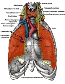
The nerve supply ( innervation ) of the diaphragm is provided by the diaphragmatic nerve ( nervus phrenicus ). In humans, this arises from the third to fifth neck segment (C3 to C5) of the spinal cord and is part of the cervical plexus ( plexus cervicalis ). In addition, so-called secondary phrenici ( Nervi phrenici accessorii ) can occur, i.e. additional nerves from the neck segments C5 to C7. In most mammals, all parts of the phrenic nerve come from the fifth to seventh neck segment, so the phrenic nerve in animals corresponds to the minor phrenic nerve in humans. A small proportion of the diaphragmatic muscles is also innervated by branches of the spinal nerves of the posterior breast segments.
The central control of the diaphragm takes place via the respiratory center in the elongated medulla and in the bridge . From here the motor root cells of the phrenic nerve in the cervical marrow are rhythmically stimulated independently of the will. The diaphragm is thus partially subject to autonomous control . Like the rest of the skeletal muscles, the diaphragm can also be influenced at will. This takes place via nerve tracts from the cerebral cortex , which, for example, enable you to “hold your breath”.
The blood supply takes place through several smaller arteries . The arteria pericardiacophrenica (only in humans) and the arteria musculophrenica (all mammals) arise from the internal thoracic artery ( arteria thoracica interna ). In addition, from the thoracic part of the aorta in humans the superior phrenic artery (upper diaphragmatic artery ) and from the abdominal part of the aorta in all mammals the inferior phrenic artery (lower diaphragmatic artery , referred to in animals as the posterior diaphragmatic artery, caudal artery ) to the Diaphragm. In some mammals (e.g. predators , pigs ) the anterior abdominal artery ( arteria abdominalis cranialis ) also supplies the diaphragm. The blood drains through the veins of the same name .
Emergence
In the embryo , the body cavity is initially undivided and is referred to here as an intraembryonic coelom . The diaphragm arises initially in the front neck area from four different systems .
The tendon plate arises from the transverse septum . This is a partition that grows out of the mesoderm and grows from the stomach side towards the back, but without reaching it. The so-called pleuroperitoneal membranes ( Membranae pleuroperitoneales ) grow from the back wall of the coelom towards the transverse septum . The lumbar part of the diaphragm arises from the back mesentery of the esophagus ( mesoesophagus dorsalis ). The section in the angle between the ribs and the diaphragm ( Recessus costodiaphragmaticus ) arises from the body wall itself. Muscle precursor cells ( myoblasts ) of the myotomes of the neck migrate into the later muscular parts , which explains the predominant innervation from branches of the cervical nerves.
In its further development, with the stretching of the neck and the displacement of the heart in the embryo, the diaphragm experiences a shift in the caudal direction (in humans downwards, in animals accordingly backwards) into its final position.
function
The diaphragm is the "motor" of the so-called diaphragmatic breathing (less aptly also called abdominal breathing ). The role of the diaphragm during inhalation ( inspiration ) is supported by other muscles, the so-called inspiratory muscles, which, by lifting the ribs, contribute to a further enlargement of the chest ( chest breathing ). The ratio of chest to diaphragmatic breathing varies according to the species as well as age, training and load-dependent and can also be arbitrarily influenced. In infants and the elderly, diaphragmatic breathing dominates, in adults it moves 60 to 80% of the air inhaled. In most mammals, chest breathing can maintain adequate ventilation of the lungs for rest and low stress, even with complete paralysis of the diaphragm (diaphragmatic palsy ).
The diaphragm , which is curved towards the chest, contracts when you inhale. In humans it is shortened by a maximum of 30 to 34%. During this contraction it flattens out and the dome shape changes into a cone shape. The firm connection with the inferior vena cava at the apex of the diaphragmatic dome contributes to this change in shape, the vena cava hole shifts only slightly downwards and forwards (in animals correspondingly backwards and downwards, technically caudoventrally ). In addition, the contraction of the diaphragm also causes the lower edge of the ribs to rise slightly and thus also a certain expansion of the chest. Above all, the diaphragmatic action expands the space in the angle between the chest wall and the diaphragm, the costodiaphragmatic recess .
This process enlarges the chest cavity and thus increases the negative pressure in the hermetically sealed pleural cavity. In most mammals, the pleural lining of the lungs ( pleura pulmonalis ) is adhesively connected to the inner lining (also made of pleura) of the chest cavity and thus also to the diaphragm by a film of liquid, the so-called pleural fluid . The lungs and the inner wall of the chest cavity / diaphragm are connected like two moistened glass panes placed on top of one another, which, although they cannot be lifted from one another, can be moved against one another. Since this fluid cannot expand, as the chest cavity increases, so does the lungs. Now flows with open glottis air into the lungs, since the outer air pressure is greater than the pressure in the lungs. In some mammals ( elephants , tapirs ) the two pleural leaves are fused together, here the lungs naturally have to follow the expansion of the chest cavity.
The contraction of the diaphragm displaces the organs of the upper abdomen (or the anterior abdominal cavity in animals) downwards (or backwards). By slackening the abdominal muscles and bulging the abdominal wall, the organs are provided with the necessary space so that there is no pressure increase in the abdominal cavity during normal breathing. Since the abdominal movement is only a passive consequence, the term "diaphragmatic breathing" should be preferred to "abdominal breathing".
When you breathe out ( exhalation ), the diaphragm relaxes. The elastic fibers in the lungs and the surface forces in the alveoli (retraction forces) cause the lungs to contract and the diaphragm to return to its dome shape. During breathing, exhalation takes place at rest, i.e. without active involvement of muscles.
In addition to the respiratory function, the diaphragm can be used together with the abdominal muscles to build up pressure in the abdominal cavity, namely when they contract at the same time, so the bulging of the abdomen is prevented. This takes place e.g. As when bowel movements or contractions instead. In exhalation techniques such as breathing support, the diaphragm works together with the rest of the respiratory muscles. The involvement of the diaphragm in laughing has already been pointed out in the section “Word meanings”.
The lumbar part of the diaphragm acts as an “external sphincter” and supports the lower esophageal sphincter , a complex locking mechanism at the transition from the esophagus to the stomach . The contraction of the diaphragm, by narrowing the esophageal slit, leads to an increase in pressure in the esophageal sphincter muscle with every inhalation.
Finally, the changes in pressure within the chest cavity caused by the diaphragm are important for the transport of blood in the veins of the chest cavity.
- Training of the diaphragm muscles
In sport, the diaphragm muscles have a double function: on the one hand, they are particularly challenged by intensive breathing, on the other hand, the tense muscles help stabilize the upper body. Even if breathing exercises (also at the open window) were part of the standard training program for middle and long distance runners up to the 1930s, they were later neglected. But since the diaphragm muscles can be trained in a similar way to other muscles, it is necessary to strengthen the muscles through strength exercises. For example, training against water resistance is useful for this. Research on this shows that with appropriate training the fatigue residues of the respiratory muscles decrease significantly.
Dysfunctions and diseases
A cramp of the diaphragm leads to - usually harmless - hiccups ( singultus ). This is a clonic spasm . According to the more recent opinion, the also mostly harmless side stitches are caused by cramping of the diaphragm as a result of an insufficient supply of oxygen. Persistent and therefore life-threatening diaphragmatic cramps can occur , for example, in tetanus .
As a result of increased pressures in one of the two body cavities, the position of the diaphragm may change. An increased pressure in the abdominal cavity leads to an elevated diaphragm , for example with liver enlargement , spleen enlargement , stomach overload , tumors or pregnancy . A low diaphragm can result from obstructive pulmonary disease or a pleural effusion . With these changes in position of the diaphragm, breathing is restricted.
An injury to the diaphragm, for example a hole ( perforation ) with a sharp object or a rupture of the diaphragm , can have life-threatening consequences if it cannot create negative pressure in the chest cavity ( pneumothorax ). In the case of foreign body disease in ruminants, foreign bodies from the reticulum perforate the diaphragm and can cause purulent pericardial inflammation or pneumonia , but disorders of the respiratory mechanics practically never occur.
As diaphragmatic hernia ( diaphragmatic hernia ) is referred to a passage of abdominal organs by the diaphragm into the chest cavity. A diaphragmatic hernia can be congenital due to a malformation of the diaphragm; it is observed in 1 in 2,500 newborns. The contents of the abdominal cavity can protrude into the pleural cavity, the mediastinum or even into the pericardial cavity ( peritoneopericardial diaphragmatic hernia , the most common congenital hernia in dogs and cats ). After birth, diaphragmatic hernias are mostly traumatic ( accidents , heavy lifting, pregnancy , overweight , sport ). A distinction is made between real diaphragmatic hernias, in which a bulging of the peritoneum surrounds the herniated organs, and diaphragmatic ruptures, which occur without the formation of a hernial sac made of peritoneum. Hernias occur most often at the weak points of the diaphragm (Bochdalek gap, Morgagni hole and Larrey gap) or in the area of the aortic or esophageal slit ( hiatal hernia ). A hiatal hernia favors the reflux ( reflux ) of acidic stomach contents into the esophagus, since the diaphragm can then not contribute to the locking mechanism of the lower esophageal sphincter.
In the case of hyperventilation syndrome , a psychological disorder leads to an increase in the frequency of contraction ("diaphragmatic neurosis").
An inflammation of the diaphragm is as Diaphragmitis referred. It is a localized muscle inflammation ( myositis ), which is usually associated with elevated diaphragm, pain and restricted movement of the diaphragm. Muscle inflammation that is limited to the diaphragm is extremely rare. A common cause was the infestation with trichinae ( trichinellosis ). This parasitic disease has largely been suppressed by the legally prescribed trichinae test for all slaughtered animals that are not pure herbivores . During the trichinae examination, a muscle sample is taken from the loin part of the diaphragm (pillar of the diaphragm) of the slaughtered animal and examined microscopically for the presence of trichinae.
Damage to the diaphragmatic nerve ( phrenic paralysis ) or paraplegia above the origin of the diaphragmatic nerve and thus an interruption between the respiratory center and the phrenic root cells leads to paralysis of the diaphragm ( diaphragmatic paralysis or diaphragmatic palsy ). Diaphragmatic palsy can also occur in the context of a so-called neuralgic shoulder amyatrophy with failure of parts of the brachial plexus or an internal capsule syndrome, which is often based on a circulatory disorder in the lenticulostriate arteries. Unilateral damage to the nerve leads to a paradoxical movement of the diaphragm ( Kienböck sign ): The diaphragm moves upwards on the diseased side due to the negative pressure during inhalation and downwards due to the increase in pressure during exhalation. A unilateral transection of the diaphragmatic nerve ( phrenicotomy ) was previously used to immobilize a lung, e.g. B. performed in tuberculosis ( Ferdinand Sauerbruch was one of the pioneers in the field of diaphragmatic surgery before the First World War ). More dangerous than phrenic paralysis are ( generalized ) disorders of the neuromuscular conduction affecting the whole body (e.g. in the case of myasthenic crisis , botulism or due to curare and other muscle relaxants ) because they affect the entire respiratory muscles and lead to respiratory arrest .
"Diaphragm" of the crocodiles
Crocodiles have a capsule of connective tissue around the liver, which forms a partition between the chest and abdominal cavity. It corresponds most closely to the transverse septum (see above) of mammals, since the liver in mammals also arises in the transverse septum and in adults it is closely connected to the diaphragm by short liver ligaments .
The paired diaphragmatic muscle ( Musculus diaphragmaticus ) of the crocodiles arises on both sides of the pelvis and the inside of the last rib. The muscle pulls forward in a fan-shaped widening and mostly attaches to the liver capsule. Some fibers run medially to the pericardium . The diaphragmatic muscle is most closely comparable to the loin part of mammals, although the diaphragm and diaphragmatic muscle of crocodiles are not homologous . During the contraction, both diaphragmatic muscles shift the liver backwards, so the liver, like the piston of a pump (" liver piston mechanism", English hepatic piston ), enlarges the chest cavity and thus the lungs. This special breathing mechanism is responsible for the fact that the crocodiles hardly ever notice any external breathing movements.
Diaphragm as a food
The cattle's diaphragmatic pillars are called kidney cones in the kitchen language , the remaining muscle parts are called hem or crown meat .
literature
- Margot A. van Herwaarden, Melvin Samsom, André JPM Smout: The role of hiatus hernia in gastro-oesophageal reflux disease. In: European journal of gastroenterology & hepatology. Vol. 16, No. 9, 2004, ISSN 0954-691X , pp. 831-835, PMID 15316404 .
- Rainer Klinke , Hans-Christian Pape , Stefan Silbernagl (eds.): Physiology. 5th, completely revised edition. Thieme, Stuttgart et al. 2005, ISBN 3-13-796005-3 .
- Christian Plathow, Christian Fink, Sebastian Ley, Michael Puderbach, Monica Eichinger, Astrid Schmähl, Hans-Ulrich Kauczor: Measurement of diaphragmatic length during the breathing cycle by dynamic MRI, comparison between healthy adults and patients with an intrathoracic tumor. In: European radiology. Vol. 14, No. 8, 2004, ISSN 0938-7994 , pp. 1392-1399, PMID 15127220 , ( digital version (PDF; 320 KB) ).
- Anat Ratnovsky, David Elad: Anatomical model of the human trunk for analysis of respiratory muscles mechanics. In: Respiratory physiology & neurobiology. Vol. 148, No. 3, 2005, ISSN 1569-9048 , pp. 245-262, PMID 16143282 , doi : 10.1016 / j . or 2004.12.016 .
- Gert-Horst Schumacher , Gerhard Aumüller : Topographical anatomy of humans. 7th edition. Urban & Fischer, Munich et al. 2004, ISBN 3-437-41367-8 .
- Franz-Viktor Salomon, Hans Geyer, Uwe Gille (ed.): Anatomy for veterinary medicine. Enke, Stuttgart 2004, ISBN 3-8304-1007-7 .
Web links
- Breathing in alligators (English)
Individual evidence
- ↑ Duden spelling: diaphragm
- ↑ Luis-Alfonso Arráez-Aybar, José-L. Bueno-López, Nicolas Raio: Toledo School of Translators and their influence on anatomical terminology . In: Annals of Anatomy - Anatomischer Anzeiger . tape 198 (3) , 2015, pp. 21–33 , doi : 10.1016 / j.aanat.2014.12.003 ( elsevier.com [accessed April 19, 2019]).
- ↑ Arnd Krüger : Many roads lead to Olympia. The changes in training systems for medium and long distance runners (1850–1997). In: Norbert Gissel (Hrsg.): Sports performance in change (= writings of the German Association for Sports Science. 94). Czwalina, Hamburg 1998, ISBN 3-88020-322-9 , pp. 41-56.
- ↑ Toshiyuki Ohya, Ryo Yamanaka, Masahiro Hagiwara, Marie Oriishi, Yasuhiro Suzuki: The 400- and 800-m Track Running Induces Inspiratory Muscle Fatigue in Trained Female Middle-Distance Runners. In: Journal of strength and conditioning research. Vol. 30, No. 5, 2016, pp. 1433-1437, PMID 26422611 , doi : 10.1519 / JSC.0000000000001220 .
- ↑ Arnd Krüger: diaphragm training. In: competitive sport. Vol. 32, No. 4, 2002, p. 36.
- ↑ Scott K. Poweers, R. Andrew Shanely: Exercise-induced changes in diaphragmatic bioenergetic and antioxidant capacity. In: Exercise and Sport Sciences Reviews . Vol. 30, No. 2, 2002, pp. 69-74, ( online ).
- ↑ Heinz-Walter Delank: Neurology. 5th, revised and supplemented edition. Enke, Stuttgart 1988, ISBN 3-432-89915-7 , p. 63 f. ( Etiopathogenesis of the plexus lesions ) and 75 f. ( Inner capsule syndrome ).
- ^ Ferdinand Sauerbruch, Hans Rudolf Berndorff : That was my life. Kindler & Schiermeyer, Bad Wörishofen 1951; cited: Licensed edition for Bertelsmann Lesering, Gütersloh 1956, p. 167.


