Guinea pig diseases
Guinea pig diseases are health disorders in the Caviidae family . In principle, house guinea pigs are susceptible to the same diseases as their wild form, but house guinea pigs have a number of other diseases that are primarily due to feeding or husbandry. In the case of feeding errors in domestic guinea pigs, a diet that is too high in carbohydrates , fat or protein and low in raw fiber is a common cause of a multitude of problems, which can range from dental diseases, diarrhea, bloating, urinary stones to cramps and even death of the animal . Posture errors such as poor hygiene, wet litter, drafts or direct sunlight or too high ambient temperatures also promote the occurrence of a number of diseases.
Digestive system disorders
The guinea pig is adapted to a low-energy, fiber-rich diet. It has teeth that grow back for life ( rootless teeth ), which require constant tooth abrasion through intensive chewing and gnawing activity. The smooth muscles of the gastrointestinal tract are poorly developed. The ingestion of many small meals and fiber are important for the intestinal peristalsis and thus for the further transport of the food in the gastrointestinal tract. The difficult to digest food is broken down by bacteria in the large intestine . Feeding errors quickly lead to disturbances in the delicate balance of this intestinal flora . Like humans, guinea pigs cannot produce vitamin C in the body, so they are dependent on its ingestion through food. The food of guinea pigs should therefore consist almost or better exclusively of hay and fresh, not too cold, green fodder (see also Guinea pig nutrition ).
More common signs of a digestive problem are reduced or absent food intake ( anorexia ), diarrhea and flatulence (" drum addiction ").
Teeth that are too long lead to feeding problems. In addition to insufficient gnawing opportunities, possible causes are also congenital or acquired tooth misalignments and traumatic tooth loss. A lack of tooth abrasion can promote bridging of the molars , which prevent the tongue from lifting up to the roof of the mouth and thus make swallowing impossible, or lead to a pike bite . Since the teeth are no longer used, a vicious circle sets in. Typical signs of a dental problem are anorexia and increased salivation with damp fur around the mouth. The disturbed food intake quickly causes degeneration of the chewing muscles and serious diseases of the gastrointestinal tract. In addition, the animals often try to compensate for the lack of food by drinking more ( polydipsia ) or preferring soft food. Dental problems have to be corrected by shortening the incisors or grinding the bridges, usually at regular intervals, as the permanent growth causes tooth misalignments to occur again and again. Damage in the area of the incisor root can lead to the formation of a " giant tooth " ( macrodont ), which must be shortened regularly.
Diarrhea is almost always due to disturbances in the intestinal flora as a result of incorrect feeding or the use of the wrong antibiotics , which favor secondary infections of the intestine. Antibiotics with a spectrum of action on only gram-positive bacteria (e.g. penicillin ) lead to a strong increase in gram-negative bacteria and thus to antibiotic-associated diarrhea or antibiotic-related enterotoxemia . Poor postural hygiene also promotes the occurrence of intestinal diseases. Here in guinea pigs the Hefepilzerkrankung dominate Darmmykose by Cyniclomyces guttulatus (obsolete Saccharomyces or Saccharomycopsis guttulatus ) or pintolopsii Torulopsis , something rarely occur secondary bacterial diseases such as salmonellosis ( Salmonella typhimurium , S. enteritidis ) and infections caused by Clostridium difficile -related (especially through the use wrong antibiotics), Escherichia coli , Yersinia pseudotuberculosis , Pseudomonas aeruginosa , Listeria monocytogenes or non-specific germs such as streptococci and staphylococci . In addition, there are individual case reports on the occurrence of Tyzzer's Disease ( Clostridium piliforme pathogen ), which is usually fatal. Viral intestinal inflammations are occasionally caused by coronaviruses with weaning and are fatal in 50% of the animals. Also, the infection of the colon with nematodes (especially Paraspidodera uncinata , a Pfriemenschwanz ) occasionally in outdoor entertainment before and can lead to diarrhea, but remains mostly without clinical symptoms. More rarely, single-celled organisms cause diarrhea. The coccidiosis ( Eimeria caviae ) may result in pups in larger conversations bloody diarrhea, wherein here also secondary intestinal mycoses or bacterial intestinal inflammation play a role. More rarely, trichomoniasis ( Tritrichomonas caviae , T. flagellipora ), amoeba dysentery ( Entamoeba caviae ) or an infection with Cryptosporidium wrairi also cause diarrhea. For treatment, feeding errors must first be corrected. Antimycotics (e.g. nystatin ) are also used for intestinal fungal infections, antibiotics for bacterial infections, toltrazuril or sulfonamides for coccidia , metronidazole or fenbendazole for other single-cell diseases . In addition, forced feeding with diet food, the administration of probiotics to stabilize the natural intestinal flora and, in the case of severe diarrhea, a fluid replacement are indicated.
Flatulence manifests itself in a distended belly and mostly occurs as a result of feeding errors or reduced food intake (teeth). In guinea pigs, constipation occurs less often, the longer retention time of the food pulp in the gastrointestinal tract tends to lead to incorrect fermentation and thus to colon (intestinal symptoms) or gastric inflation ( gastric symptoms ), and rarely to gastric torsion . This inflation is very painful (teeth grinding, reduced food intake, fatigue ) and can lead to circulatory failure ( shock ) or to the affected part of the digestive tract bursting. Both conditions are therefore emergencies. In the case of lighter forms, treatment is carried out by administering drugs that stimulate intestinal motor skills ( metoclopramide ) and foam-breaking drugs ( dimeticone ). If the tympanum is severe, it may be necessary to insert a gastric tube or puncture the inflated appendix and to administer antibiotics and painkillers. Subsequently, errors in feeding and, if necessary, the teeth must be corrected and force feeding with dietary feed must be carried out.
In diseases of the liver , fatty liver disease is common with a high- energy diet. This usually remains for a long time without clinical symptoms and only becomes acute when there is a lack of energy and manifests itself unspecifically in loss of appetite and fatigue, in severe cases in loss of consciousness ( hepatic coma ) and cramps . Infectious liver inflammation ( hepatitis ) occurs in guinea pigs with general infections with streptococci or staphylococci as well as with Tyzzer's disease. When subjected to external violence, traumatic hepatitis can develop if the liver is injured.
Respiratory diseases
Respiratory diseases manifest themselves in shortness of breath ( dyspnea ) and increased breathing noises , in infections also in nasal discharge and often also conjunctivitis ( conjunctivitis ) with eye discharge.
In guinea pigs, respiratory diseases are very often bacterial, the discharge is then purulent. Pasteurella multocida ( Pasteurellosis ), Bordetella bronchiseptica , Klebsiella pneumoniae , Chlamydophila caviae , Streptobacillus moniliformis , as well as strepto- and staphylococci and various Haemophilus species play a role as pathogens . Respiratory infections are treated with antibiotics and agents that dissolve secretions ( acetylcysteine , bromhexine ). The chances of recovery from pneumonia in guinea pigs are rather poor compared to other animals.
Viral respiratory infections are also relatively common . According to Ewringmann and Glöckner, guinea pig-specific adenovirus pneumonia is quite common, according to Wasel, at least in mass poses. In a more recent study, 1% of the guinea pigs sent for dissection were diagnosed with adenovirus pneumonia. Apparently it does not require any secondary bacterial infection. It has an incubation period of 5 to 10 days and manifests itself in the death of the airway mucosa as a result of bronchiolitis and is usually fatal. According to Huerkamp et al. Adenoviral pneumonia is rare and viral respiratory infections are more frequently caused by paramyxoviruses ( Sendai virus , Simian 5 virus and Murine pneumonia virus ), especially when they come into contact with mice and rats .
Serious trauma (falls, dog bites) can lead to pulmonary bleeding with red, frothy blood coming out of the nose. Pulmonary hemorrhage is an emergency that requires immediate veterinary attention. Heart disease ( hypertrophic cardiomyopathy with pulmonary edema or pleural effusion ) and extensive space-occupying processes in the abdomen such as inflation of the stomach or intestines also cause breathing problems. Pathogens that have got into the blood ( septicemia ), especially in intestinal diseases, can settle in the respiratory system. Tumorous neoplasms in the area of the lungs (lung adenomas ) are relatively common. Allergic respiratory diseases, on the other hand, are rare in guinea pigs.
Diseases of the urinary organs
Diseases of the urinary organs are manifested by a change in the properties of the urine or in the frequency of urination. Guinea pigs ingest calcium from their diet in an unregulated manner, and excess calcium is excreted in the urine. Adequate fluid intake is required for this. Due to the alkaline pH value of the urine (around 8.5), physiologically calcium crystals precipitate, which is why the urine of the guinea pig is yellowish to whitish cloudy. Due to the phytochemicals excreted via the kidneys and oxidation of urine components, the urine can also turn reddish in the air, which should not be confused with blood.
Due to the peculiarity of calcium metabolism, guinea pigs tend to develop urinary stones ( urolithiasis ). In guinea pigs, around 85% of the stones consist entirely or predominantly of calcium carbonate , mostly in the form of calcite , and more rarely vaterite . These calcium carbonate stones often have a calcium phosphate content, so they are mixed stones. For bladder infections due to Ureasebildnern also struvite occur when conditions in oxalic acid rich feeding and calcium oxalate . They lead to irritation of the mucous membrane of the urinary tract and possibly also to secondary bacterial infection by ascending germs, which can lead to an inflammation of the bladder ( cystitis ) in particular . Cystitis is most common in females over 2 years old. The result is pain when urinating ( stranguria ) and frequent urination ( pollakiuria ), possibly with blood. The anal region is then often wet and there is a risk of fly maggot infestation ( myiasis ). The stones can also lead to the obstruction of the urinary tract, whereupon the formation of urine (in the case of renal pelvic and ureteral stones) as well as the urination (in the case of bladder and urethral stones) is no longer possible ( anuria ). If the urinary bladder outlet or the urethra are obstructed, the bladder fills to the point of bursting (emergency).
Reproductive system disorders can cause discharge or cystitis-like symptoms.
Increases in scope
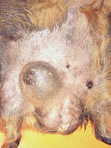
Local increases in size are common in guinea pigs. Local encapsulated pus foci ( abscesses ) occur mainly on the lower jaw as a result of dental diseases, on the other parts of the body after bite injuries or from impaled food. A second common cause of increased circumference are groats ( atheromas ), which can be the size of a hen's egg and occur mainly in the area of the back, the flanks and the caudal gland . Both abscesses and atheromas must be treated surgically, simply expressing the contents is usually not sufficient as they refill.
Older guinea pigs can also develop benign fatty growths ( lipomas ) that do not require treatment. They make up about 40% of skin tumors in guinea pigs. The second most common skin tumor with a share of 30% is the hair follicle tumor ( trichofolliculoma ). Malignant tumors such as fibrosarcomas (around 10% of skin tumors) or bone tumors ( osteosarcomas ) are significantly less common, and treatment is usually futile for the latter.
Swelling of the lymph nodes in the head and neck area in guinea pigs is mainly caused by the bacterium Streptococcus zooepidemicus , which penetrates the body through small injuries. The lymph node swelling caused by an oncornavirus is less common in leukemia , whereby the lymph nodes in other parts of the body ( inguinal lymph nodes , knee fold lymph nodes ) are usually also affected.
Mammary tumors occur in guinea pigs of both sexes. About three quarters are benign adenomas, the remainder are malignant adenocarcinomas . They show up as an increase in circumference in the groin region , in which the two mammary complexes (one on each side of the body) of the mammary gland are located. Swelling of the breasts in older females who have been pregnant several times can also be caused by harmless gynecomastia . In pregnant females, inflammation of the mammary gland ( mastitis ) occurs with a frequency of about 2% , which is mainly caused by coli bacteria and treated with broad spectrum antibiotics (e.g. chloramphenicol ).
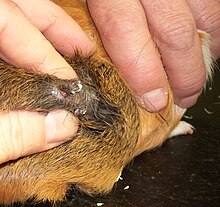
A thickening in the area of the perineum can be caused by a blockage of the perineal pocket , a skin pocket between the anus and the genital opening into which the sebum glands open. Constipation occurs mainly in older males and can also be caused by too soft faeces (e.g. in intestinal mycosis). In this case, the perineal pockets filled with feces, sebum and epithelial residues must be emptied manually and treated with mild skin disinfectants. In the perineal region, large clumps of hair, litter, feces and urine can also develop in the event of diarrhea or the need to urinate. Like clogged perianal pockets, these are often used by flies to lay their eggs and fly maggot infestation (myiasis) occurs. A hard increase in circumference in front of the genital opening is usually a urinary stone stuck in the urethra .
If there is swelling in the throat area, an enlargement of the thyroid gland ( goiter ) must also be considered.
Skin diseases

Skin diseases are expressed in skin ( efflorescences ) and / or coat changes. These can be primarily caused by the disease, or secondary, especially scratching as a result of itching . Since guinea pigs scratch their back legs, skin changes when itching occurs mostly in the front part of the body.
The most common cause of skin changes in guinea pigs is infestation with ectoparasites . The guinea pig mange (pathogen Trixacarus caviae ) as well as infestation with fur mites ( Chirodiscoides caviae ) and with hair lice ( Gyropus ovalis , Gliricola porcelli , Trimenopon hispidum ) dominate. The latter two can be recognized with the naked eye; the microscopic examination of a skin scrap is required to detect Trixacarus caviae . Infestation with other skin parasites is much less common. There are no lice and fleas specifically adapted to guinea pigs , but if they come into contact with other rodents, mouse and rat lice ( Polypax serrata , Polypax spinulosa ) or dog and cat fleas can migrate to guinea pigs. Demodicosis ( Demodex caviae ) or Cheyletiellosis ( Cheyletiella parasitovorax ) are very rare . Occasionally, allergic reactions to storage and food mites (e.g. Acarus farris ) also occur. The skin parasitoses are treated with ectoparasitics such as fipronil , ivermectin , doramectin , selamectin or amitraz . The leishmaniasis seen in ulcerative skin lesions, but is only in South America.
In second place after skin parasites are bacterial dermatitis and skin fungal diseases ( dermatomycosis and dermatophytosis ), the former also after previous damage by parasites and scratches. A bacterial skin inflammation is characterized by greasy deposits and treatment is carried out with antibiotics. The main skin fungus in guinea pigs is Trichophyton mentagrophytes , which primarily leads to dandruff and hair loss in the back area. Antifungal drugs are used for treatment.
Frequent causes of skin wounds are bite injuries, if kept in groups by other animals, but also by rabbits, dogs and cats. It is not uncommon for vaccination reactions to occur even after subcutaneous injections , which lead to dry death and rejection of an area of the skin, but are otherwise harmless and heal on their own.
In addition, skin diseases caused by hormonal imbalances also occur in guinea pigs. In females, the most common cause is estrogen- active ovarian cysts , which usually cause symmetrical hair loss in the flank. There is usually no itching in these animals.
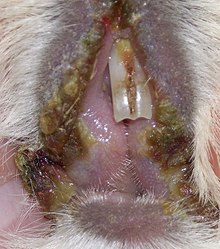
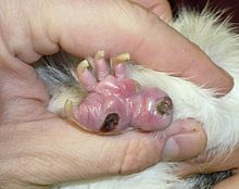
There are special skin diseases in three locations. The lip Grind ( Cheylitis ) results in scabs on the mouth, the cause of this disease is so far unclear which treatment is therefore difficult. At the base of the tail, the guinea pig has a special skin gland , the caudal gland. This can lead to inflammation or tumors (adenomas), which show up in thickenings, adhesions and reddening. Tumors should be surgically removed as early as possible. The sole ulcer occurs mainly in overweight animals, with long claws and poor housing conditions. Here it comes to inflammation of the ball, in severe cases to severe general disorders and severe lameness, which then often only allow euthanasia.
Reproductive system diseases
Female animals
In female guinea pigs often occur cysts of the ovary to (ovarian cysts). The cause of this has not yet been clarified; it is probably a hormonal disorder . The cysts can become very large and, due to the mass, impede bowel movements or breathing and cause the abdomen to swell. Hormonally active cysts are usually small and produce large amounts of estrogens . This can lead to symmetrical hair loss in the flank, weight loss and a weakening of the immune system and thus to an increased susceptibility to infections. Large cysts can be punctured through the abdominal wall . Hormonal treatment with hCG then delayed again formation of cysts for some time. The only curative treatment is surgical removal of the ovaries ( ovariectomy ), which is associated with a considerably higher risk of anesthesia in guinea pigs than in dogs, cats or rabbits.
Another relatively common condition in females is thickening of the uterine lining . In contrast to domestic dogs , guinea pigs rarely develop an accumulation of pus in the uterus ( pyometra ), but rather an accumulation of blood ( hemometra ). This blood is usually released in spurts with the urine, so that the disease can be mistaken for a urinary bladder infection or urinary stones. Tumors of the uterus can also lead to bleeding. Leiomyomas , more rarely adenocarcinomas , occur in guinea pigs . In the cervix , there may be a thickening of the mucous membrane with an increase in the mucous glands there, which is known as endocervical hyperplasia .
Birth disorders occasionally occur in guinea pigs. Too early breeding use (before the 6th month of life), late first breeding use (after the 11th month of life), dead fetuses or a mostly feeding-related calcium deficiency, which leads to weakness in labor , are beneficial .
Male animals
In male guinea pigs, secretion stones (concretions) can form in the seminal vesicle glands . The general condition is not disturbed, however symptoms similar to urinary bladder inflammation can occur. The formation of secretion stones is relatively rare and occurs in less than 1% of the animals.
Occasionally, deck active bucks develop a chronic inflammation of the penile foreskin and glans ( balanoposthitis ), which is caused by foreign bodies that have penetrated the foreskin cavity after copulation. If it persists for a long time, it can lead to paraphimosis .
Hormonal imbalances
Diabetes ( diabetes mellitus ) is rare in guinea pigs. The cause has not yet been researched. It is not uncommon for it to be a laboratory diagnostic incidental finding and the animals are symptom-free. Other animals eat and drink a lot, urinate frequently and tend to become obese. Opacity of the lens of the eye occurs relatively often as a concomitant phenomenon. In advanced cases, liver damage can also occur and the animals are knocked off or comatose . Treatment with long-acting insulin preparations should only be carried out in the case of pronounced clinical symptoms and repeated blood glucose checks.
With the relatively frequent overactive thyroid gland ( hyperthyroidism ), the animals drink and urinate a lot and lose weight. The appetite is increased at the beginning, but in the further course the feed consumption decreases. Greasy diarrhea and a tendency to develop throat infections due to damage to the immune system are also observed. Hair loss often occurs, usually beginning in the groin area . If the thyroid is greatly enlarged, a swelling in the neck area may be visible.
Increased water intake (polydipsia) is not only observed in hormonal disorders, but can also occur in dental diseases as well as postural (boredom, behavioral disorders) and feeding (little green fodder). The administration of glucocorticoids used as anti-inflammatory agents also leads to increased fluid intake.
Skeletal diseases
Broken bones (fractures) occur in guinea pigs mainly after kicking or falling. The bones of guinea pigs are prone to splintering. The thighbone is particularly often affected. Fractures lead to lameness with relief of the affected limb. There is also a risk of inflammation of the bones (osteomyelitis). Fractures usually have to be treated surgically ( osteosynthesis : intramedullary nailing or external fixator ).
Malignant bone tumors ( osteosarcomas ) can occur in older guinea pigs, especially on the upper arm bone . The animals show increasing lameness and pain. A treatment is not useful in most cases, a euthanasia recommended.
Satin guinea pigs suffer from a special bone disease, osteodystrophy , which is associated with limb pain and attempts to relieve pressure ("tinkling"). This hereditary disease cannot be treated.
Joint diseases also occur in guinea pigs. Acute inflammation ( arthritis ) usually occurs after injuries or sole ulcers and must be treated with antibiotics. Chronic changes in joints ( arthrosis ) can appear as a sign of wear and tear in older animals and treatment is usually limited to the administration of pain reliever medication.
Neurological diseases
Neurological diseases are rather rare in guinea pigs. The most common cause is trauma, which leads to damage to the spinal cord or, in the case of head injuries, to the brain . The result is mostly paralysis , with severe damage the prognosis is poor. Guinea pig paralysis, which occurs rarely, must also be taken into account in the case of symptoms of paralysis. The causative agent of this disease of the spinal cord is not yet known. Guinea pig paralysis cannot be treated and usually leads to the death of the animal within 10 days. Paralysis can also enjoy a stroke , a meningitis , an inflammation of the brain , brain abscesses or brain tumors occur.
Head tilts are mostly caused by an inflammation of the inner ear resulting from an otitis media . Middle ear infections (pathogens Streptococcus equi , Bordetella bronchiseptica , Streptococcus pneumoniae , Escherichia coli , Klebsiella or pseudomonads ) are relatively common (incidence 15%), but usually remain symptom-free. It only spreads to the inner ear in about 1% of cases. Although 25 to 50% of the guinea pigs had antibodies against Encephalitozoon cuniculi in various studies , no clinical diseases have been documented after infection with this pathogen.
In guinea pigs, seizures are usually caused by guinea pig mange, as the animals suffer from extreme itching when the infection is severe. Typically, these attacks can be triggered by vigorous stroking. Liver, kidney, heart and brain diseases, anemia , septicemia, heat stroke , poisoning ( oleander ) and hypoglycaemia can also cause attacks. In overweight females one week before to one week after giving birth, ketosis (“pregnancy toxicosis”) must be considered. This is usually peracute and can therefore no longer be treated.
Eye diseases
Guinea pigs have an extensive body of fat in their eye socket . In some breeds or over-nourished animals, “fat eyes” can develop , with the lower eyelid bulging outwards (apparent ectropion ). This is not a disease and does not require treatment.
A cataract , i.e. clouding of the lens of the eye , occurs in guinea pigs for hereditary reasons, as a symptom of old age or in the presence of diabetes. Cataract treatment is not common in guinea pigs. Lens changes such as cataracts or sclerosis of the eye lens are the most common eye diseases in guinea pigs, they occur in around 38% of animals.
Conjunctivitis (conjunctivitis) with reddening of the conjunctiva and eye discharge is mostly caused by foreign bodies (hay dust) or drafts, but it can also be a concomitant disease with respiratory infections (see above). Conjunctivitis is also typical with a severe vitamin C deficiency. Foreign objects can also cause injury to the cornea and inflammation of the cornea ( keratitis ).
A special eye disease in guinea pigs is osseous choristia . This leads to bone formation in the ciliary body , which shows up as whitish, clasp-like opacities in the periphery of the visible part of the eyeball. The cause of the disease is unknown and it usually does not affect the animal. In exceptional cases, it can lead to inflammation of the middle skin of the eye ( uveitis ) or glaucoma ( glaucoma ).
Occasionally there is also an inflammation of the tear sac ( dacryocystitis ), which usually originates from the tips of the tooth roots.
literature
- W. Beck, N. Pantchev: Parasitoses of the guinea pig . In: Practical Parasitology in Pets . Schlütersche Verlagsgesellschaft, Hanover 2006, pp. 35-60, ISBN 3-89993-017-7 .
- A. Ewringmann, B. Glöckner: Key symptoms in guinea pigs, chinchilla and degu . Enke Verlag, Stuttgart 2005, ISBN 3-8304-1055-7 .
- I. Hamel: The guinea pig as a patient . Enke Verlag, Stuttgart 2002, ISBN 3-8304-1002-6 .
- MJ Huerkamp et al .: Guinea pigs . In: K. Laber-Laird et al .: Handbook of rodent and rabbit medicine . Pergamon Press, 1996, pp. 91-149, ISBN 0-08-042505-4 .
- O'Rourke: Disease problems of guinea pigs . In: KE Quesenberry, JW Carpenter: Ferrets, rabbits and rodents. Clinical medicine and surgery . 2nd ed., Saunders, St Louis 2004, pp. 245-254, ISBN 0-7216-9377-6 .
- E. Wasel: guinea pigs . In: K. Gabrisch, P. Zwart: Diseases of pets . 6th edition, Schlütersche Verlagsgesellschaft, Hanover 2005, pp. 49–86, ISBN 3-89993-010-X .
- Doris Quinten, Frank Malkusch: Guinea pig diseases . Ulmer, Regensburg 2007, ISBN 978-3-8001-5454-8 .
Web links
- Urinary stones in guinea pigs. Retrieved December 13, 2014 .
Individual evidence
- ↑ Birgit Drescher, Ilse Hamel: Guinea pigs: pet and patient pet and patient . Georg Thieme Verlag, 3rd edition 2012, ISBN 9783830411581 , p. 15.
- ↑ a b c d C.-C. Sommerey et al .: Diseases of the guinea pigs from the point of view of pathology . Veterinarian Praxis Kleintiere 32 (2004), pp. 377–383.
- ^ Urinary Stone Analysis Center Bonn: Animal Stone Letter 1/2010.
- ↑ a b Pet tumors. In: Laboklin aktuell 4/2009
- ↑ a b Anja Ewrigmann and B. Glöckner: Diseases of the urinary and genital organs in rodents and rabbits. In: Tierärztliche Praxis Volume 36, 2008, Suppl. 1, pp. S78 – S82
- ↑ Christof A. Bergmann et al .: Increase in the circumference of the cervix uteri in a guinea pig . In: Kleintierpraxis Volume 65, 2020, Issue 7, pp. 400–403.
- ↑ Peng, X et al .: Cystitis, urolithiasis and cystic calculi in aging guineapigs . Lab Anim 1990; 24: 159-63.
- ↑ Anja Ewringmann, Barbara Glöckner: Key symptoms in guinea pigs, chinchilla and degu . 2nd Edition. Georg Thieme Verlag, 2012, Stuttgart 2012.
- ↑ Barbara L. Oglesbee: Blackwell's Five-Minute Veterinary Consult: Small Mammal . John Wiley & Sons, 2011, ISBN 978-0-8138-2018-7 , pp. 303 .
- ↑ William D. Sullivan: Ocular disease in the guinea pig (Cavia porcellus): A survey of 1000 animals. In: Vet. Ophthalmol. 13 (2010), pp. 54-62.


