
Endometriosis
| Classification according to ICD-10 | |
|---|---|
| N80 | Endometriosis |
| N80.0 | Endometriosis of the uterus (adenomyosis uteri) |
| N80.1 | Endometriosis of the ovary |
| N80.2 | Endometriosis of the uterine tuba |
| N80.3 | Endometriosis of the pelvic peritoneum |
| N80.4 | Endometriosis of the rectovaginal septum and vagina |
| N80.5 | Endometriosis of the intestine |
| N80.6 | Endometriosis in a scar |
| ICD-10 online (WHO version 2019) | |
Endometriosis (new formation from the endometrium , ' uterine lining ', with the suffix ancient Greek ωσις = -osis for diseases, the former from ancient ἔνδον endon 'inside' and μήτρα mētra 'uterus') is a common, benign , often painful chronic disease in women in which the lining of the uterus (the endometrium) is similar to tissue outside the uterine cavity ( ectopic ).
Like the lining of the uterus , endometrial tissue changes during the menstrual cycle and can cause tissue bleeding, scarring, and pain. Endometriosis usually occurs in the lower abdominal or pelvic area , for example on the uterus or the fallopian tubes. The ovaries are often affected. An attack on the uterine muscles is called adenomyosis (adenomyosis uteri) . It spreads to the vaginal wall and the intestine (large intestine). Endometriosis can also occur in the lungs or in other internal organs such as B. occur in the brain , even if this is rarely the case. Endometriosis can be a cause of infertility in women. The cause of the development of endometriosis is not yet medically known. In addition, there is no possibility of prophylaxis for the clinical picture. Endometriosis is primarily treated surgically by removing or destroying the affected lesions. Secondary hormonal inhibition of the cycle can be used to reduce the likelihood of endometriosis recurrence . In a widely distributed, diffuse endometriosis in the uterine wall (adenomyosis) is even a hysterectomy indicated and there is no other therapy at the time.
Epidemiology
frequency
Endometriosis is the second most common gynecological disease after myomas . Around 4 to 12 percent of all women develop it between puberty and menopause , in Germany around 40,000 each year. Exact information on the frequency ( prevalence and incidence ) does not exist. The prevalence differs according to the clinical situation. For women with menstrual pain, it is 40 to 60 percent, for women with chronic abdominal pain it is over 30 percent and for women with an unfulfilled desire to have children it is around 20 to 30 percent. The disease was also found in first-degree relatives (mothers, sisters) of endometriosis patients, with a prevalence of 6.9 percent, compared with only 1 percent of the husbands' mothers and sisters. For gynecological operations through an abdominal incision is found in 1 to 15 percent, in all gynecological laparoscopy endometriosis (laparoscopy) 5 to 53 percent and laparoscopies of Fertility in patients 30 to 50 percent. Only half of the women concerned show symptoms .
Diagnosis
Endometriosis is a hormone-dependent disease that is extremely rare in prepubertal girls and rarely in postmenopausal women. It is most commonly diagnosed in women between the ages of 25 and 35. The mean age at diagnosis was 28 years. However, the diagnosis can sometimes be made just a few months after the first menstrual period , in individual cases even before the first menstrual period.
| Time to diagnosis | |
|---|---|
| Women < 19 years | 12.1 years |
| Women > 30 years | 3.0 years |
| Patients who wish to have children | 4.0 years |
| Patients with pelvic problems | 7.0 years |
Endometriosis is often diagnosed late. On average, 3 to 11 years elapse between the first symptoms and the diagnosis. During this time, around 50 percent of the women concerned visited five or more doctors. The younger the patient is at the first signs, the more time it will take for endometriosis to be diagnosed.
The reasons for the delayed diagnosis are the unspecific symptoms , which, like menstrual cramps ( dysmenorrhea ), are sometimes viewed as normal by girls, women and doctors. Therefore complaints are reported with a delay and sometimes misinterpreted by doctors, whereby a large number of differential diagnoses are possible causes of complaints.
Non-specific treatments such as the administration of hormonal contraceptives can alleviate the symptoms and thus also delay the correct diagnosis. Ultrasound examinations are used as the only examination for pelvic complaints, although only endometriosis cysts of the ovaries can be reliably detected with them.
localization


Endometriosis is usually found on the peritoneum in the lower abdomen or in the small pelvis . The Douglas space , the ligaments between the uterus and sacrum and the ovaries are often affected. Spread to the vaginal wall and the intestine and the connective tissue between the vagina and intestine are not uncommon. The disease is also found in the uterine muscles , the appendix and the diaphragm . The vulva , perineum, and navel can also be affected. Endometriosis very rarely affects the lungs , skeletal muscles and the brain .
In the abdomen, a distinction is made between superficial peritoneal endometriosis, which only affects the peritoneum, and so-called deep infiltrating endometriosis, which grows into structures such as the urinary bladder and intestinal wall or ureter .
Endometriosis can also be found in scars after operations , for example after caesarean sections .
| infested structure | Frequency of infestation in percent |
|---|---|
| Sacrouterine ligament | 60 |
| Ovaries | 52 |
| Douglas room | 28 |
| bladder | 15th |
| Ligamentum latum | 16 |
| Rectum | 12 |
| Mesosalpinx | 10 |
| Ligamentum rotundum | 5 |
| Fallopian tubes | 2 to 8 |
| Appendix vermiformis | 2 |
Emergence

causes
The causes of the development of endometriosis are not fully understood. There are numerous explanatory models, but they have not yet been proven. The transplant theory by Sampson assumes that loose endometrial cells in the retrograde menstruation through the fallopian tubes, but also about the blood and the lymphatic vessels as well as in operations deported to and settle elsewhere. In the Metaplasietheorie by Meyer arise endometriosis in place of embryonic peritoneal cavity cells , the so-called coelomic epithelium . The induction theory is a combination of the transplantation and metaplasia theory. Genetic factors also seem to play a role in the disease. Environmental toxins such as PCB , DDT or dioxins can, among other things, have a similar effect to estrogens and thus possibly disrupt the endocrine system during the prenatal phase. Immunological causes are also discussed. The Archimetratheorie of Leyendecker sees the endometriosis as a result of adenomyosis or their early manifestations and thus as a disease of archimetra on. This concept is based on specific changes in normal endometrium in women with endometriosis, to archimetrale dysfunctions such as hyper- and dys peristalsis and a archimetrale infiltration into the uterine muscle (myometrium). The Archimetra concept was further developed by Leyendecker into the Tissue Injury And Repair Theory (TIAR). After that, increased peristalsis of the uterine muscles leads to the smallest injuries in them (autotraumatisation). In the repair mechanism, estrogens are released locally, which in turn increase peristalsis. This could create a vicious circle . However, there is also evidence that there is a connection with increased estrogens for adenomyosis, but not for other endometriosis localizations.
However, none of the theories presented so far can explain the complex picture of endometriosis. Rather, one has to adopt a multimodal concept from the previously known theories, in which a large number of different factors interact. Weinschenk saw a disorder of the autonomic nervous system as the trigger for the faulty peristalsis.
| Theory / cause | description | supporting documents |
|---|---|---|
| Transplant theory |
|
|
| Metaplasia theory |
|
|
| Induction theory |
|
|
| genetics |
|
|
| Environmental toxins |
|
|
| Immunological causes |
|
|
| Tissue Injury And Repair Theory (TIAR) |
|
|
| Concept of autonomic nervous system malfunction |
|
|
| Cell and molecular biology concepts |
|
|
| Aromatase concept |
|
|
Risk factors
The main risk factor is menstruation itself. Women with an early onset of menstruation and a late menopause are at increased risk. Women who suppress their menstrual period by constantly taking hormonal contraceptives have a lower risk of developing endometriosis. Women with a short cycle less than 28 days and bleeding regularly for more than seven days were found to be at increased risk. Women whose family members are already sick also show a higher incidence of the disease. Late first pregnancy , interventions on the uterus and obesity are further risk factors . However, a connection with a low body mass index and body fat distribution is also discussed.
Symptoms
The main symptoms of endometriosis are cramp-like pain of increasing intensity associated with the menstrual cycle ( dysmenorrhea ), but chronic abdominal and back pain also occur. Some scientists regard dysmenorrhea as an early symptom of endometriosis rather than a consequence. If the Douglas area is affected , pain during sexual intercourse or painful or difficult bowel movements ( dyschezia ) can occur. Similarly, pain when urinating described. The period pains correlate neither with the size of the endometrial foci nor with a special anatomical location. The emergence of permanent pain is associated with a strong expansion of the herd. It is assumed that deeply penetrating endometrial islands irritate the smallest nerves in the pelvic space either by compression or by infiltration. The menstrual period can be increased and prolonged. A urinary obstruction may indicate a deep infiltrating endometriosis. In around half of the patients, however, the disease causes little or no symptoms.
| Symptoms | Frequency in percent |
|---|---|
| Dysmenorrhea | about 95 |
| Lower abdominal pain | approx. 85 |
| nausea | approx. 82 |
| Bowel symptoms | approx. 78 |
| prolonged and heavy menstruation | approx. 65 |
| Discomfort during intercourse ( dyspareunia ) | approx. 60 |
| Headache , dizziness | n / A |
| Stomach discomfort | n / A |
| unwanted childlessness | n / A |
| frequent infections | n / A |
| subfebrile temperatures | n / A |
Endometriosis-related sterility
The fertility (fecundability) of healthy women is between 0.15 and 0.20 successful pregnancies per month and decreases with age. In untreated women with endometriosis, the fecundability is between 0.02 and 0.10 and thus below that of healthy people. It is estimated that 30 to 50% of women with endometriosis are sterile (infertile). So far, however, no causal connection between endometriosis and sterility has been identified if there are no organic changes in the area of the fallopian tubes and ovaries. However, endometriosis is often associated with sterility. Mechanical factors due to adhesions in the fallopian tubes are accepted as the cause. A too fast and irregular transport of sperm due to the increased uterine peristalsis and too high intrauterine pressure can prevent fertilization and implantation of the egg cell. Other factors can include impaired ovarian and fallopian tube function, impaired follicle growth , premature conversion of unbroken follicles into yellow bodies ( luteinized unruptured follicle syndrome , also LUF syndrome ), hyperprolactinemia , corpus luteum insufficiency , immunological dysregulation, autoantibodies and a changed composition of the abdominal cavity fluid with increased activated macrophages , cytokines , prostaglandins . Surgical removal of endometrial foci in women who wish to have children increases the likelihood of spontaneous pregnancy . If endometriosis recurs, especially after several surgical interventions, artificial insemination is superior to another operation to fulfill the desire to have children.
Endometriosis and Pregnancy
Before drugs such as GnRH analogs were available, pregnancy was considered a natural treatment for endometriosis. Pregnancy is now only considered a possible corrective factor for the further development of endometriosis. Amenorrhea lasting 9 months and possibly also a prolonged absence of menstrual bleeding due to breastfeeding (lactation amenorrhea ) occurs. If the pregnancy is carried to term, there is a pronounced decidualization of endometriosis and, after the birth , apoptosis . Endometriosis cannot be cured by pregnancy. The data from the Nurses' Health Study II showed that women who breastfed longer reduced their risk of endometriosis.
On the other hand, endometriosis seems to have a negative impact on pregnancy, as women with endometriosis are more likely to develop preeclampsia, bleeding during pregnancy, premature placenta detachment, placenta previa, or premature rupture of the bladder. The risk of complications in childhood, such as premature birth or growth retardation, is increased.
Endometriosis in men
Usually men do not get endometriosis. Treatment with high doses of estrogen for prostate cancer can, however, lead to endometriosis in individual cases, which is seen as evidence of the metaplasia theory of the development of endometriosis.
Endometriosis in animals

Adenomyosis has been described in various mammals such as baboons , brazzamats , chimpanzees , orangutans , rhesus monkeys , dogs , cats, and mice . However, endometriosis at other locations appears to be limited to primates .
After a caesarean section, primates are at risk of endometriosis in the abdominal cavity due to scattered endometrial fragments. Animal experiments have shown adenomyosis in the uterine scar after caesarean sections in sheep .
Impaired fertility has been found in baboons associated with endometriosis . They are therefore considered to be a good animal model for research into the disease and the associated fertility problems in humans. Rhesus monkeys and mice are used as further animal models.
The in horses and various other domestic species known disease endometrosis , a chronic - degenerative disease of the endometrium , is not related to endometriosis.
pathology
Endometriosis is a benign condition. Due to the different behavior and different findings during the tissue examination, endometriosis is divided into different subtypes:
- the endometriosis of the peritoneum
- the endometriosis of the ovaries
- the deep infiltrating endometriosis
- the adenomyosis
In addition, a distinction is made between active and inactive endometrial foci based on clinical, tissue and, if necessary, biochemical aspects.
| Macroscopy | |||
|
Peritoneal endometriosis active red foci vesicular deposits |
Peritoneal endometriosis inactive black, brown foci, whitish peritoneal thickenings |
Ovarian endometriosis active thin-walled cysts bluish, bleeding in, red |
Ovarian endometriosis inactive thick-walled cysts fibrotic , brown, black |
| microscopy | |||
|
Epithelium active endometrioid highly differentiated hormonal changes eutrophic many mitoses |
Epithelium inactive, narrowed, cube-shaped, low differentiation, no hormonal changes, atrophic some / no mitoses |
Stroma active edema , inflammation erythrocytes many capillaries eutrophic - |
Stroma inactive condensed, fibrosis siderophages few / no capillaries condensed - |
| biochemistry | |||
| Detection of antibodies against the body's own (autologous) endometrium | |||
Clinical appearance
In the case of endometriosis on the peritoneum, a distinction is made between red, white and black foci or between pigmented and non-pigmented (atypical) foci. The red and unpigmented foci are considered early forms of endometriosis. They are considered to be particularly active because they show pronounced signs of cell proliferation . The typical blue-black foci, mostly with a white border, are rather old and mostly no longer active findings.

Blue-black foci with whitish pitted changes
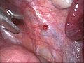
Red endometrial focus

Unpigmented white stove
Endometriosis of the right ovary


In the uterine muscles, endometriosis stimulates hyperplasia of the muscles, so that the wall thickness of the uterus increases. In addition, small cystic cavities form. Because of this reaction, the disease is called adenomyosis here . Endometrial foci can also occur in myomas , which are then called adenomyomas .
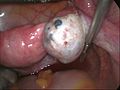
Cystic cavities also form in the ovaries over a longer period of time. These are called endometriomas or, according to their content (thickened old blood), as tar or chocolate cysts . This is actually a superficial endometriosis, in which an endometriosis foci initially forms on the surface, which gradually invades into the ovary.

Opened endometriosis cyst of the left ovary

Endometriosis on the peritoneum between the bladder and uterus

Massive adhesions in endometriosis

Adenomyosis uteri


In deep infiltrating endometriosis, pronounced nodular changes are found, especially in the area of the ligaments, such as the sacrouterine ligament, and in the rectovaginal space between the vagina and rectum. These changes can grow into surrounding organs such as the urinary bladder and rectum, as well as deep into the retroperitoneum . Pronounced retroperitoneal endometriosis can lead to a narrowing of the ureters with congestion of the urine and even hydronephrosis .
Microscopic picture
Histologically, there are small islands of tissue that consist of stroma and enclose typical endometrial glands . The glandular tissue can show hormonal changes as in the normal uterine lining. Also, can hemosiderin -loaded macrophages , known siderophages find.

Endometrioma in a microscopic image

Endometriosis of the ovary in a microscopic image

Adenomyosis uteri in the microscopic image


.JPG)
In adenomyosis, the endometrial stroma and glands are embedded in the smooth muscles of the uterine wall.
Smooth and striated muscle cells , along with stroma and glands, can be found in the foci in deep infiltrating endometriosis. These foci show pronounced proliferative properties, but only minor hormonal changes. A research group from the Johns Hopkins University School of Medicine in Baltimore reported somatic mutations in the cells of the lesions in the majority of women after exome analysis of foci of deeply infiltrating endometriosis in 24 women.
Malignant endometriosis-associated diseases
The change from endometriosis to malignant disease is possible, but extremely rare and then usually affects the ovaries. They are often recognized earlier than other ovarian cancers and usually show a higher degree of differentiation than these. You will then be treated like ovarian cancer, assuming a worse response to chemotherapy .
The most common locations outside the genitals are the rectum , the sigmoid colon and the rectovaginal septum. The therapy here consists of resection, if possible in a healthy state, and subsequent radiation therapy .
In the context of menopausal hormone replacement therapy , estrogen monotherapy in endometriosis patients is a potential risk factor for the occurrence of such malignant diseases and is therefore not recommended.
classification
The classification of endometriosis is handled very inconsistently. Many different schemes have been developed in order to enable comparability of the characteristics of the findings. The localization, the extent or the size of the herd and the endometriosis-related consequential damage are usually used as criteria. So far, none of the staging can meet all requirements. In particular, there is no correspondence between the stage and the patient's existing symptoms.
Classification according to localization
According to the localization of the foci, endometriosis is divided into three forms:
- Endometriosis genitalis interna: Endometriosis foci within the myometrium (also adenomyosis uteri )
- External genital endometriosis: foci of endometriosis outside the uterus in the pelvis (ovaries, sacrouterine ligament, Douglas, bladder peritoneum)
- Endometriosis extragenitalis: foci of endometriosis outside the pelvis
Staging
The most widespread today is the classification of the American Society for Reproductive Medicine (so-called rASRM or earlier AFS staging). It has established itself internationally and works with a point system, according to which a distinction is made between four stages / degrees of severity from easy to severe , as well as the additional description of the activity of the endometrial lesions. However, it does not take into account extragenital forms of the disease, as does the older EEC (Endoscopic Endometriosis Classification) stage classification, which goes back to Kurt Semm and is based on a purely descriptive assessment. It too is divided into four stages. The WHO classification corresponds to this staging. Since the description of retroperitoneal and deep infiltrating growth forms of endometriosis is not sufficient in all stages used so far, the ENZIAN classification was developed. In analogy to the oncological staging of the TNM or FIGO system , she tries to divide the disease into four degrees of severity. The demarcation of the stages results from the extent of the disease in relation to the area and the depth. To assess the chances of pregnancy in endometriosis was Endometriosis fertility index (EFI) developed which, in addition to endometriosis classification according rASRM, the functional limitations, the woman's age, duration of infertility involving and previous pregnancies in the analysis. In the STCDAP classification (Subtle-Typical-Cystic-Deep-Adenomyosis-Pocckets), delicate, typical and cystic foci, deep endometriosis, adenomyosis and peritoneal pockets are classified separately.
| WHO / EEC | rASRM | GENTIAN | EFI | |
| criteria |
|
|
|
|
| Inclusion of extragenital findings | Yes | No | Yes | No |
| Assessment of deep infiltrating endometriosis | No | No | Yes | No |
| Assessment of the activity | No | only by macroscopic description of the herd | No | No |
| Assessment of the chances of pregnancy | No | No | No | Yes |
diagnosis
The suspicion that endometriosis may be present often arises after a precise anamnesis of the occurrence and nature of the pain as well as diseases of close relatives.
A large number of diseases from the fields of gynecology , gastroenterology and urology must be included in the differential diagnosis .
| Gynecological diseases | Gastroenterological diseases | Urological diseases |
|---|---|---|
|
Manual examination of the pelvis (vaginal, rectal, rectovaginal) can only reveal larger endometrial foci. Foci on the vulva , in the vagina and on the external cervix can be viewed colposcopically .

A transvaginal ultrasound scan can reveal large foci of endometriosis. If the rectum is suspected, an additional transrectal ultrasound examination can be helpful. Magnetic resonance imaging can help clarify the extent of a deep infiltrating endometriosis in individual cases . However, this cannot rule out the disease if the imaging tests are negative.
The determination of CA-125 in the blood does not make sense as a diagnosis due to insufficient specificity and is only suitable to a limited extent for monitoring the progress.
Endometriosis of the abdominal cavity can only be demonstrated with a laparoscopy . The foci are assessed visually and partially or completely removed for tissue protection.
The method published in 2009 of examining uterine lining samples for certain nerve fibers that occur only or only in large quantities in women with endometriosis is currently not an established diagnostic method. In 2011, British researchers described profiles of proteins in the urine that could possibly be used once to diagnose and assess the severity of the disease.
therapy
The aim of endometriosis treatment is to improve the quality of life by eliminating pain, eliminating dysfunction of affected organs and successfully treating infertility (sterility, infertility). A distinction is made between symptomatic treatment ( pain treatment ), surgical and drug therapy and the use of complementary forms of therapy.
Operative therapy


The aim of the surgical treatment is to remove as much as possible all endometrial foci by cutting out ( extirpation ) or to destroy them safely with heat ( electrocautery , laser surgery ) and to restore the anatomical conditions as far as possible. Chocolate cysts in the ovaries should be peeled off while preserving the ovaries. In addition, any adhesions that may be present can be loosened and, if you wish to have children, the patency of the fallopian tubes can be checked by means of chromopertubation . The procedure can be performed with a laparoscopy or, especially with extensive findings, with an abdominal incision . Especially with superficial endometriosis foci, laparoscopic surgical therapy is a very effective form of treatment. Repeated surgical interventions, on the other hand, can result in organ loss (e.g. ovaries) and iatrogenically cause additional pain through scarring and adhesions.
The therapy of choice for symptomatic deep infiltrating endometriosis is complete removal of the foci. This can be done via vaginal resection, laparoscopy, laparoscopically assisted vaginal surgery or an abdominal incision. If you want to have children, the necessary preservation of the uterus may only allow an incomplete resection of the endometriosis.
The pain and prolonged or increased bleeding caused by diffuse endometriosis in the uterine muscles (adenomyosis uteri) can be successfully treated with a removal of the uterus if there are no other causes of symptoms. Individual adenomyomas can be surgically removed.
There is no unanimous opinion among experts regarding the effects of the surgical treatment of endometriosis on the desire to have children. There seems to be a small benefit for laparoscopic rehabilitation of peritoneal endometriosis. Rectovaginal endometriosis excision appears to be of dubious use in improving fertility. The excision of ovarian endometriomas appears to have the greatest benefit in terms of pregnancy rate.
| ESHRE 2005 | |
| Grade I / II endometriosis | limited use, surgery recommended |
| Grade III / IV endometriosis | possible but unproven benefit, surgery recommended |
| Endometrial removal before IVF | recommended if the endometrioma is> 4 cm |
| Recurrent endometriosis | no recommendations |
| ASRM 2004 | |
| Grade I / II endometriosis | little benefit, surgery recommended |
| Grade III / IV endometriosis | possible benefit, surgery recommended |
| Endometrial removal before IVF | dubious benefit, no recommendations |
| Recurrent endometriosis | not recommended |
| RCOG 2006 | |
| Grade I / II endometriosis | proven benefit, surgery recommended |
| Grade III / IV endometriosis | possible benefit, recommendation uncertain |
| Endometrial removal before IVF | recommended if the endometrioma is> 4 cm |
| Recurrent endometriosis | no recommendations |
Medical therapy
Surgical measures to secure the suspected diagnosis, especially if you want to have children and if endometrioma is suspected, are the first choice in therapy. Medicinal treatment strategies can also be useful as an alternative, but also as a long-term concept, to alleviate symptoms or prevent the disease from recurring. A fundamental distinction is to be made between symptomatic treatment of pain and therapy that has a direct or indirect effect on endometriosis.
Pain relievers such as acetylsalicylic acid , ibuprofen , diclofenac , naproxen or indomethacin are used to treat the pain .
An effect on the endometriosis can be achieved hormonally through single-phase estrogen-progestogen combination preparations (“ pill ”), pure progestins such as medroxyprogesterone acetate , cyproterone acetate , chlormadinone acetate , levonorgestrel and dienogest , as well as GnRH analogues . The testosterone derivative danazol is also effective in the treatment of endometriosis, but was withdrawn from the market in Germany in 2005 due to an unfavorable risk-benefit ratio.
If taken continuously without a break ("long cycle"), estrogen-progestin combinations lead to regression of the uterine lining and a significant reduction in pain in endometriosis. However, there is no detailed evidence of the effects on the herds.
With continuous use, pure gestagens lead to a blockage of the ovarian function by inhibiting the hypothalamus - pituitary - ovary axis and, in the optimal case, to amenorrhea . This leads to an improvement in the pain, a regression or inhibition of growth or the formation of new endometriosis. They can also be given in the form of a minipill , a 3-month syringe or an IUD .
GnRH analogues also suppress the hypothalamus-pituitary-ovary axis and thus lead to a lowering of the estradiol level with a lack of menstruation after the first 4 weeks. Typical climacteric symptoms such as hot flashes and sleep disorders occur. A significant reduction in bone density ( osteoporosis ) occurs with long-term treatment. By means of a so-called add-back treatment with the accompanying administration of gestagens or estrogen-progestin combinations, the symptoms and the loss of bone mass can be compensated without measurably influencing the effectiveness of the endometriosis treatment. The duration of the GnRH analog treatment is limited to about six months. It is currently the most effective type of drug treatment and, in higher stages, also leads to an increase in the success rate of fertility treatments. For ovarian endometriomas, drug treatment alone is not sufficient. An administration of GnRH analogues prior to surgery can lead to a reduction in the size of the endometrioma. Whether this will make the operation easier or reduce the recurrence rate is a matter of controversy in the specialist literature. GnRH analogues after surgery on an endometriosis cyst do not compensate for incomplete surgery. Likewise, the benefit of pre- or postoperative GnRH analog therapy for deeply infiltrating endometriosis has not been proven.
Treatment with GnRH antagonists, aromatase inhibitors , COX-2 inhibitors , selective progesterone receptor modulators and estrogen receptor β agonists for the treatment of endometriosis is currently still being tested.
Complementary treatment
Complementary treatment approaches can support surgical and drug therapy. However, there is no evidence-based evidence of their effectiveness. As part of order therapy, more physical exercise should be encouraged and relaxation processes and meditation techniques should be taught. Exercise therapy, massage , mud baths and yoga are considered good measures for symptomatic treatment of dysmenorrhea.
Changing your diet may help alleviate the suffering. It should help to avoid milk, dairy products and wheat, to eat a lot of fresh fruit and vegetables, cold-pressed oils such as evening primrose oil, linseed oil, olive oil, and fatty sea fish ( omega-3 fatty acids ).
Further treatment options are physical , enzyme , microbiological and phytotherapy , acupuncture , neural therapy and traditional Chinese medicine . Obesity should be reduced and tobacco smoking given up.
forecast
Endometriosis is a chronic disease with a high rate of recurrence . Regardless of the original treatment, be it surgical rehabilitation, drug suppression of ovarian function or a combination of surgical and drug treatment, stage-dependent recurrence rates between 20 and 80% have been reported. Relapses cannot be prevented, especially after drug treatment has been discontinued. They can only be postponed for the period of hormonal treatment. A three- to six-month drug treatment with GnRH analogues also increased after surgery the chances of fertility treatment , but only in stages III and IV (after rASRM). There is no unanimous opinion among experts regarding the benefit of surgical treatment of endometriosis for fulfilling the desire to have children.
history
The oldest known description of the disease is the medical doctoral thesis "Disputatio Inauguralis Medica de Ulceribus Ulceri" by the German doctor Daniel Shroen from 1690. In it he described "ulcers" that become significant in the entire abdomen, between the bladder, bowel and ligamentum latum Lead adhesions.
In 1860, Carl von Rokitansky (1804–1878) described the disease and established it in modern medicine. Because of the common occurrence of uterine lining and smooth muscles, he called the findings adenomyomas . In 1889 Hermann Löhlein reported his observation of an "adenomatous disease of the corpus uteri with multiple cyst formation in the corpus wall" to the Society for Obstetrics and Gynecology in Berlin. For many years, adenomyomas and bleeding ovarian cysts were thought to be different clinical pictures.
The Canadian gynecologist Thomas Stephen Cullen (1868-1953) described endometrial foci of the ligamentum rotundum in 1896 and recognized the common cause of the clinical pictures. Johannes Pfannenstiel (1862–1909) was the first to describe endometriosis in the rectovaginal septum in 1897 .
The German gynecopathologist Robert Meyer described scar endometriosis in 1903 and endometriosis in the sigmoid colon , a section of the large intestine , and in lymph nodes in 1909 . In 1919 he developed the metaplasia theory of the development of the disease.
John A. Sampson (1873–1946) developed the transplant theory in 1921 and the term endometriosis in 1927 . The Austrian Oskar Frankl introduced the term adenomyosis in 1925 and explained in 1929 that an endometriosis, which he called adenomyosis tubae , also occurs in the fallopian tubes . He ruled out an inflammatory cause for the thickening of the fallopian tubes near the uterus, which up until then had been assumed and the phenomenon was therefore called salpingitis isthmica nodosa . The term endometriosis , coined by Sampson, only finally caught on until 1932.
Economical meaning
Endometriosis is of considerable economic importance due to medical expenses and lost work . The costs associated with endometriosis were in the 2002 United States 22 billion dollars estimated. This is offset by costs of $ 865 million for Crohn's disease and $ 13 billion to $ 17 billion for migraines . According to the results of a British study, patients are unable to work an average of 45 days a year due to significant complaints. However, women with endometriosis, regardless of absence from work, have a significantly reduced productivity in their working life due to pain. In addition, they are impaired by pain in everyday activities. On the other hand, endometriosis causes little or no discomfort in around half of the patients.
Despite the high number of an estimated 40,000 newly diagnosed women in Germany each year and its associated high economic importance, the disease has so far been relatively little represented in clinical and basic scientific research.
Organizations
Various organizations have been founded internationally to raise awareness of the disease and to support research and further training.
The World Endometriosis Society (WES) was founded in 1998 and promotes the exchange of knowledge between scientists from different disciplines. She regularly hosts the World Congresses on Endometriosis .
To promote research in the field of endometriosis, the World Endometriosis Research Foundation (WERF) was founded in 2006 , in which the Special Interest Group for Endometriosis & Endometrium (SIGEE) of the European Society of Human Reproduction and Embryology (ESHRE), the Endometriosis Special Interest Group (EndoSIG) of the American Society for Reproductive Medicine (ASRM) and the World Endometriosis Society .
In September 2005 the European Endometriosis League (EEL) was founded. Affiliated with the EEL is the Endometriosis Research Foundation (SEF), which supports the work of the EEL by organizing joint meetings, seminars, training events and congresses on the subjects of endometriosis, pain and infertility. The endometriosis congress of German-speaking countries takes place every two years.
Together with the Endometriose-Vereinigung-Deutschland e. V., a Germany - wide self-help organization , the EEL and the SEF certify endometriosis centers to improve the diagnosis and treatment of the disease.
Every year in the second week of March, an international Endometriosis Awareness Week takes place to draw attention to the disease.
literature
Specialist literature
- Journal of Endometriosis , ISSN 2036-282X , Important Editore Verlag, Milan ( Italy )
- Thomas Steck, Ricardo Felberbaum, Wolfgang Küpker, Cosima Brucker, Dominique Finas: Endometriosis . Springer, 2004, ISBN 3-211-00746-6 ( limited preview in Google book search).
- Andreas D. Ebert: Endometriosis: A guide for practice. Walter de Gruyter, 2006, ISBN 3-11-018984-4 ( limited preview in the Google book search).
- Liselotte Mettler : Endometriosis. pmi Verlag, Frankfurt am Main, 2000, ISBN 3-89786-022-8 .
- RW Shaw: An Atlas of Endometriosis. Parthenon Publishing Group, Carnforth-Pearl River 1993.
- Gülden Halis among others: Diagnosis and therapy of deeply infiltrating endometriosis . In: Dtsch Arztebl Int . No. 107 (25) , 2010, pp. 446-455 ( Article ).
Guidelines
- S2k guidelines for endometriosis: Diagnostics and therapy of the German Society for Gynecology and Obstetrics (DGGG). In: AWMF online (as of 8/2013)
- ESHRE guideline for the diagnosis and treatment of endometriosis. of the European Society of Human Reproduction and Embryology . In: Hum Reprod. 20, 2005, pp. 2698-2704, doi: 10.1093 / humrep / dei135 , guidelines.endometriosis.org
Lay literature
- Ewald Becherer, Adolf Schindler: Endometriosis: Advice and help for those affected and their families. W. Kohlhammer Verlag, 2009, ISBN 978-3-17-020342-6 .
- Nicole von Hoerschelmann: Endometriosis - Pain-free through optimal nutrition and a healthy handling of stress. Diametric Verlag, 2011, ISBN 978-3-938580-21-9 .
- Jörg Keckstein , Christiane Niehues, Anja Engelsing, Ansgar Römer, Karl W. Schweppe, Hans R. Tinneberg: Endometriosis - the misunderstood women's disease !? Diagnostics and therapy from a holistic medical perspective. Diametric Verlag, 2009, ISBN 978-3-938580-17-2 .
- Lois Jovanovic, Genell J. Subak-Sharpe: Hormones. The medical manual for women. (Original edition: Hormones. The Woman's Answerbook. Atheneum, New York 1987) From the American by Margaret Auer, Kabel, Hamburg 1989, ISBN 3-8225-0100-X , pp. 98, 112-118, 143 ff. And 373.
- Martin Sillem: Endometriosis: benign, but mean. Recognize the hidden disease and treat it effectively. TRIAS Medical Council, 2003, ISBN 3-8304-3095-7 .
- Martin Sillem: Effective help with endometriosis. A Women's Guide: How Your Doctor Treats You. TRIAS Medical Council, 1999, ISBN 3-89373-472-4 .
Web links
- Endometriosis - information at Gesundheitsinformation.de (online offer of the Institute for Quality and Efficiency in Healthcare )
- Information on endometriosis from the Gynecological Endoscopy Working Group of the German Society for Gynecology and Obstetrics (DGGG)
- Endometriose-im-Netz.de - About life with endometriosis and medical care
- Endometriosis.org - English language peer reviewed information platform
Organizations
- Endometriosis Research Foundation
- EVA. Endometriosevereinigung Austria
- Endometriosis Association Germany
- European Endometriosis League
- World Endometriosis Society
- World Endometriosis Research Foundation
- Special Interest Group for Endometriosis and Endometrial Disorders (SIGEE) of the European Society of Human Reproduction and Embryology (ESHRE)
- Endometriosis Special Interest Group (EndoSIG) of the American Society for Reproductive Medicine (ASRM)
Individual evidence
- ↑ a b c Liselotte Mettler , A. Schmutzler; In: Klaus Diedrich , Wolfgang Holzgreve, Walter Jonat , Askan Schultze-Mosgau, Klaus-Theo M. Schneider: Gynecology and Obstetrics . Springer Verlag, 2006, ISBN 3-540-32867-X ( full text in the Google book search).
- ^ Fritz Nagele: Endometriosis - An Underestimated Suffering? In: J Gynecol Endocrinol. 3, 2009, pp. 45–46, kup.at (PDF; 242 kB)
- ↑ a b A. E. Schindler, P.-A. Regidor; In: JW Dudenhausen: gynecology and obstetrics . Walter de Gruyter, 2002, ISBN 3-11-016562-7 ( full text in the Google book search).
- ↑ a b c M. S. Arruda, CA Petta, MS Abrão, CL Benetti-Pinto: Time elapsed from onset of symptoms to diagnosis of endometriosis in a cohort study of Brazilian women. In: Hum Reprod. 18, 2003, pp. 756-759. PMID 12660267 , oxfordjournals.org
- ↑ a b c d e f g h i F. Oehmke, F. Suwandinata, C. Deisting, H. Tinneberg: data on endometriosis. In: Gynecologist . 40, 2007, pp. 521-526 doi: 10.1007 / s00129-007-2015-6
- ↑ K. Yamamoto, Y. Mitsuhashi, T. Takaike, K. Takase, H. Hoshiai, K. Noda: Tubal endometriosis diagnosed within one month aftermenarche: a case report. In: Tohoku J Exp Med. 181, 1997, pp. 385-387. PMID 9163854
- ^ AD Ebert , N. Fuhr, M. David, L. Schneppel, T. Papadopoulos: Histological Confirmation of Endometriosis in a 9-Year-Old Girl Suffering from Unexplained Cyclic Pelvic Pain since Her Eighth Year of Life. In: Gynecol Obstet Invest. 67, 2008, pp. 158-161. PMID 19077389
- ^ A b H. Suginami: A reappraisal of the coelomic metaplasia theory by reviewing endometriosis occurring in unusual sites and instances. In: Am J Obstet Gynecol. 165, 1991, pp. 214-218. PMID 1853899
- ↑ RE Batt, MF Mitwally: Endometriosis from thelarche to midteens: pathogenesis and prognosis, prevention and pedagogy. In: J Pediatr Adolesc Gynecol. 16, 2003, pp. 337-347. PMID 14642954 , doi: 10.1016 / j.jpag.2003.09.008
- ↑ a b c d e f g h i j k l m Adolf E. Schindler: Epidemiology, pathogenesis and diagnosis of endometriosis. In: J Fertil Reprod. 17, 2007, pp. 22–27, kup.at (PDF; 205 kB)
- ↑ a b c d e f g h i j k l S2k guidelines for endometriosis: diagnostics and therapy of the German Society for Gynecology and Obstetrics (DGGG). In: AWMF online (as of 8/2013)
- ↑ a b c d S. Dogan, S. Djalali, A. Agic, K. Diedrich, D. Hornung: Diagnosis of endometriosis: New tests from peripheral blood. In: J Gynecol Endocrinol. 2, 2008, pp. 14–17, kup.at (PDF; 263 kB)
- ↑ a b K. W. Schweppe: Endometriosis - a disease without a lobby. In: Zentralbl Gynäkol. 125, 2003, p. 233.
- ^ GK Husby, RS Haugen, MH Moen: Diagnostic delay in women with pain and endometriosis. In: Acta Obstet Gynecol Scand. 82, 2003, pp. 649-653. PMID 12790847
- ^ S. Luisi, L. Lazzeri, V. Ciani, F. Petraglia: Endometriosis in Italy: from cost estimates to new medical treatment. In: Gynecol Endocrinol. 11, 2009, pp. 734-740. PMID 19908951
- ↑ G. Hudelist, N. Fritzer, A. Thomas, C. Niehues, P. Oppelt, D. Haas, A. Tammaa, H. Salzer: Diagnostic delay for endometriosis in Austria and Germany: causes and possible consequences. In: Hum Reprod . (2012). PMID 22990516 , doi: 10.1093 / humrep / des316
- ↑ Nina Ayerle: Severe pain - severe diagnosis: around every tenth woman is affected by endometriosis . In: Stuttgarter Nachrichten . No. 253 , October 31, 2019, p. 40 .
- ^ Ruth Hadfield, Helen Mardon, David Barlow, Stephen Kennedy: Delay in the diagnosis of endometriosis: a survey of women from the USA and the UK. In: Hum Reprod. 11, 1996, pp. 878-880. PMID 8671344 , oxfordjournals.org (PDF; 249 kB)
- ^ Karen Ballard, Karen Lowton, Jeremy Wright: What's the delay? A qualitative study of women's experiences of reaching a diagnosis of endometriosis. In: Fertil Steril . 86, 2006, pp. 1296-1301. PMID 17070183 , doi: 10.1016 / j.fertnstert.2006.04.054
- ↑ a b M. L. Ballweg: Impact of endometriosis on women's health: comparative historical data show that the earlier the onset, the more severe the disease. In: Best Pract Res Clin Obstet Gynaecol. 18, 2004, pp. 201-218. PMID 15157638
- ↑ a b K. W. Schweppe, AD Ebert, L. Kiesel: Endometriosis Centers and Quality Management. In: Gynecologist . 43, 2010, pp. 233-240, doi: 10.1007 / s00129-009-2484-x
- ↑ S. Saberi, AR Farhoud, A. Radmehr: Calf endometriosis: a case report and review of musculoskeletal involvement. In: Am J Orthop (Belle Mead NJ). 38, 2009, pp. E175-E178. PMID 20049359
- ↑ Wolfgang Kühn, Heinz Pickartz: Clinical Pathology of the female genitalia. Wissenschaftliche Verlagsanstalt, 2001, ISBN 3-8047-1778-0 , p. 223.
- ↑ a b c d e F. Wieser, R. Wenzl, RN Taylor, K. Diedrich, D. Hornung: Genetics of Endometriosis. In: Gynecologist . 37, 2004, pp. 676-680, doi: 10.1007 / s00129-004-1558-z
- ↑ a b c d e f g h W. Urdl: The current status of conservative therapy for endometriosis. In: J Reproduktionsmed Endokrinol. 3, 2006, pp. 24–30, kup.at (PDF; 429 kB)
- ↑ G. Leyendecker, G. Kunz, M. Noe, M. Herbertz, D. Beil, P. Huppert, G. Mall: The archimetra as a new morphological-functional concept of the uterus and as the location of the primary disease in endometriosis. In: Reproductive Medicine. 15, 1999, pp. 356–371, doi: 10.1007 / s004440050126 , oxfordjournals.org (PDF; 236 kB)
- ↑ G. Leyendecker, G. Kunz, M. Noe, M. Herbertz, G. Mall: Endometriosis: a dysfunction and disease of the archimetra. In: Hum Reprod Update. 4, 1998, pp. 752-762. PMID 10027630
- ^ G. Leyendecker, L. Wildt, G. Mall: Uterine peristaltic activity and the development of endometriosis. In: Ann NY Acad Sci. 1034, 2004, pp. 338-355. PMID 15731324 , PMC 2730449 (free full text)
- ↑ a b c d e f Jörg Keckstein, Gernot Hudelist, Frank Tuttlies, Uwe Ulrich: Update Endometrioseforschung. New concepts - new methods. In: gynecology + obstetrics. 2, 2010, pp. 22-25, online ( page no longer available , search in web archives ) Info: The link was automatically marked as defective. Please check the link according to the instructions and then remove this notice. (PDF; 363 kB)
- Jump up ↑ Claire Templeman, Sarah F Marshall, Giske Ursin, Pamela L. Horn-Ross, Christina A. Clarke, Mark Allen, Dennis Deapen, Argyrios Ziogas, Peggy Reynolds, Rosemary Cress, Hoda Anton-Culver, Dee West, Ronald K. Ross , Leslie Bernstein: Adenomyosis and endometriosis in the California Teachers Study. In: Fertil Steril. 90, 2008, pp. 415-424. PMID 17919609 , doi: 10.1016 / j.fertnstert.2007.06.027 , PMC 2813675 (free full text)
- ↑ a b c d e f g h Stefan Weinschenk: Endometriosis, dysmenorrhea, sterility and the autonomic nervous system. In: EHK. 53, 2004, pp. 1-9. natum.de (PDF; 1.7 MB)
- ↑ a b c d e Ingrid Gerhard, Marion Kiechle: Integrative gynecology: conventional and complementary therapy . Elsevier, Urban & Fischer, 2006, ISBN 3-437-56260-6 ( full text in the Google book search).
- ↑ a b c d e f g h i j k l m n o p q B. Hinney, G. Emons; In: Marion Kiechle: Gynecology and Obstetrics . Elsevier, Urban & Fischer-Verlag, 2006, ISBN 3-437-42406-8 ( full text in the Google book search).
- ↑ a b Grant W. Montgomery, Dale R. Nyholt, Zhen Zhen Zhao, Susan A. Treloar, Jodie N. Painter, Stacey A. Missmer, Stephen H. Kennedy, Krina T. Zondervan: The search for genes contributing to endometriosis risk . In: Hum Reprod Upd. 14, 2008, pp. 447-457. PMID 18535005 , doi: 10.1093 / humupd / dmn016
- ^ KA Hansen, KM Eyster: Genetics and genomics of endometriosis. In: Clin Obstet Gynecol. 53, 2010, pp. 403-412. PMID 20436317
- ↑ H. Cakmak, HS Taylor: Molecular mechanisms of treatment resistance in endometriosis: the role of progesterone-hox gene interactions. In: Semin Reprod Med . 28, 2010, pp. 69-74. PMID 20104430
- ^ A b c Thomas Steck, Ricardo Felberbaum, Wolfgang Küpker, Cosima Brucker, Dominique Finas: Endometriosis . Springer-Verlag, 2004, ISBN 3-211-00746-6 ( full text in the Google book search).
- ↑ a b c d Andreas D. Ebert: Endometriosis: A guide for practice . Walter de Gruyter-Verlag, 2006, ISBN 3-11-018984-4 ( full text in the Google book search).
- ↑ a b c d Cynthia Farquhar: Endometriosis. In: BMJ. 334, 2007, pp. 249-253. PMID 17272567 , doi: 10.1136 / bmj.39073.736829.BE , PMC 1790744 (free full text)
- ↑ UR Panganamamula, OH Harmanli, EF Isik-Akbay, CA Grotegut, V. Dandolu, JP Gaughan: Is prior uterine surgery a risk factor for adenomyosis? In: Obstet Gynecol. 104, 2004, pp. 1034-1038. PMID 15516398
- ↑ F. Nawroth, D. Foth, T. Schmidt, R. Sudik, Th. Römer: Differential diagnostic problems and therapy of scar endometriosis. In: obstetric women's health. 60, 2000, pp. 496-498, doi: 10.1055 / s-2000-8031
- ↑ Mary L. Hediger, Heather J. Hartnett, Germaine M. Buck Louis: Association of endometriosis with body size and figure. In: Fertil Steril. 84, 2005, pp. 1366-1374. PMID 16275231 , doi: 10.1016 / j.fertnstert.2005.05.029 , PMC 1343487 (free full text)
- ^ SE McCann, JL Freudenheim, SL Darrow, RE Batt, MA Zielezny: Endometriosis and body fat distribution. In: Obstet Gynecol. 82, 1993, pp. 545-549. PMID 8377980
- ↑ LB Signorello, BL Harlow, DW Cramer, D. Spiegelman, JA Hill: Epidemiologic determinants of endometriosis: a hospital-based case-control study. In: Ann Epidemiol. 7, 1997, pp. 267-274. PMID 9177109
- ^ S. Bérubé, S. Marcoux, R. Maheux: Characteristics related to the prevalence of minimal or mild endometriosis in infertile women. Canadian Collaborative Group on Endometriosis. In: Epidemiology. 9, 1998, pp. 504-510. PMID 9730028
- ↑ M. Kaufmann , A. Pfleiderer: Tumors and changes in the female sexual organs. In: M. Breckwoldt, M. Kaufmann , A. Pfleiderer (eds.): Gynecology and obstetrics. Stuttgart 2008, pp. 205-208.
- ↑ A. Fauconnier, C. Chapron: Endometriosis and pelvic pain: epidemiological evidence of the relationship and implications. In: Hum Reprod Update. 11, 2005, pp. 595-606. PMID 16172113 , pdf oxfordjournals.org.Retrieved December 29, 2008.
- ↑ a b c d e f g h i j R. Baumann In: Manfred Kaufmann, Serban D. Costa, Anton Scharl: Die Gynäkologie . Springer Verlag, 2005, ISBN 3-540-25664-4 ( full text in the Google book search).
- ↑ Wolfgang Janni: Specialist in gynecology . Elsevier, Urban & Fischer Verlag, 2008, ISBN 3-437-23915-5 ( full text in the Google book search).
- ^ American Society for Reproductive Medicine : Endometriosis and infertility. In: Fertil Steril. 86 (Suppl 4) (2006), pp. S156 – S160, asrm.org (PDF; 91 kB)
- ↑ U. Ulrich, F. Müller, F. Tuttlies, J. Keckstein: Diagnosis and therapy of endometriosis - current developments. In: Gynecologist. 50, 2009, pp. 506–510, online ( memento of the original from March 4, 2016 in the Internet Archive ) Info: The archive link has been inserted automatically and has not yet been checked. Please check the original and archive link according to the instructions and then remove this notice. (PDF; 275 kB)
- ↑ a b Freimut Leidenberger, Thomas Strowitzki, Olaf Ortmann (eds.): Clinical endocrinology for gynecologists . Springer Verlag, 2004, ISBN 3-540-89759-3 ( full text in the Google book search).
- ↑ Jörg Baltzer, Klaus Friese, Michael Graf, Friedrich Wolff: Practice of Gynecology and Obstetrics: The complete practical knowledge in one volume . Georg Thieme Verlag, 2006, ISBN 3-13-144261-1 ( full text in the Google book search).
- ↑ Martin Sillem: Endometriosis: benign, but mean. Recognize the hidden disease and treat it effectively. TRIAS Medical Council, 2003, ISBN 3-8304-3095-7 ( full text in the Google book search).
- ↑ Leslie V. Farland, A. Heather Eliassen, Rulla M Tamimi, Donna Spiegelman, Karin B. Michels, Stacey A. Missmer: History of breast feeding and risk of incident endometriosis: prospective cohort study. In: BMJ. 2017, p. 358, doi: 10.1136 / bmj.j3778 . PMID 28851765 , bmj.com (PDF; 476 kB)
- ↑ Janne Foss Berlac, Dorthe Hartwell, Charlotte Wessel Skovlund, Jens Langhoff-Roos, Øjvind Lidegaard: Endometriosis increases the risk of obstetrical and neonatal complications. In: Acta Obstet Gynecol Scand. Feb 9, 2017. PMID 28181672 , doi: 10.1111 / aogs.13111
- ^ Edwin Beckman, Susan Pintado, GL Leonard, WH Sternberg: Endometriosis of the prostate. In: Am J Surg Pathol. 9, 1985, pp. 374-379. PMID 2418693
- ^ JD Martin, AE Hauck: Endometriosis in the male. In: Am Surg. 51, 1985, pp. 426-430. PMID 4014886
- ↑ Ted C. Pinkert, Charles E. Catlow, Reuben Straus: Endometriosis of the urinary bladder in a man with prostatic carcinoma. In: Cancer . 43, 1979, pp. 1562-1567. PMID 445352 , doi : 10.1002 / 1097-0142 (197904) 43: 4 <1562 :: AID-CNCR2820430451> 3.0.CO; 2-W
- ↑ GR Schrodt, MO Alcorn, J. Ibanez: Endometriosis of the male urinary system: a case report. In: J Urol . 124, 1980, pp. 722-723. PMID 7452803 .
- ↑ a b Julie M. Hastings, Asgerally T. Fazleabas: A baboon model for endometriosis: implications for fertility. In: Reprod Biol Endocrinol. 4 (2006) Suppl 1, p. S7. PMID 17118171 , doi: 10.1186 / 1477-7827-4-S1-S7
- ↑ CM Moore, GB Hubbard, MM Leland, BG Dunn, RG Best: Spontaneous ovarian tumors in twelve baboons: a review of ovarian neoplasms in non-human primates. In: J Med Primatol. 32, 2003, pp. 48-56. PMID 12733602
- ↑ TM D'Hooghe, CS Bambra, BM Raeymaekers, PR Koninckx: Development of spontaneous endometriosis in baboons. In: Obstet Gynecol. 88, 1996, pp. 462-466. PMID 8752259 .
- ↑ TM D'Hooghe, CS Bambra, FJ Cornillie, M. Isahakia, PR Koninckx: Prevalence and laparoscopic appearance of spontaneous endometriosis in the baboon (Papio anubis, Papio cynocephalus). In: Biol Reprod. 45, 1991, pp. 411-416. PMID 1838282 , biolreprod.org ( Memento of the original from September 23, 2015 in the Internet Archive ) Info: The archive link has been inserted automatically and has not yet been checked. Please check the original and archive link according to the instructions and then remove this notice.
- ↑ AA Binhazim, RP Tarara, MA Suleman: Spontaneous external endometriosis in a De Brazza's monkey. In: J Comp Pathol. 101, 1989, pp. 471-474. PMID 2607018
- ↑ Breton F. Barrier, Jana Allison, Gene B. Hubbard, Edward J. Dick Jr, Kathleen M. Brasky, Danny J. Schust: Spontaneous adenomyosis in the chimpanzee (Pan troglodytes): a first report and review of the primate literature: case report. In: Hum Reprod. 22, 2007, pp. 1714-1717. PMID 17452396 , (online)
- ↑ KJ Graham, FA Hulst, L. Vogelnest, IS Fraser, CM Shilton: Uterine adenomyosis in an orangutan (Pongo abelii / pygmaeus). In: Aust Vet J. 87, 2009, pp. 66-69, 19178483.
- ↑ RM Hadfield, PL Yudkin, CL Coe, J. Scheffler, H. Uno, DH Barlow, JW Kemnitz, SH Kennedy: Risk factors for endometriosis in the rhesus monkey (Macaca mulatta): a case-control study. In: Hum Reprod Update. 3, 1997, pp. 109-115. PMID 9286735
- ↑ H. Tamada, N. Kawate, T. Inaba, M. Kuwamura, M. Maeda, T. Kajikawa, T. Sawada: Adenomyosis with severe inflammation in the uterine cervix in a dog. In: Can Vet J. 46, 2005, pp. 333-334. PMID 15943119 , PMC 1082876 (free full text)
- ^ A. Newell-Fugate, E. Lane: Intrapartum uterine rupture with coincidental uterine adenomyosis in an African wild dog (Lycaon pictus). In: J Zoo Wildl Med. 40, 2009, pp. 791-795. PMID 20063828
- ↑ J. Bulman-Fleming: A rare case of uterine adenomyosis in a Siamese cat. In: Can Vet J. 49, 2008, pp. 709-712. PMID 18827849 , PMC 2430406 (free full text)
- ↑ a b c P. Greaves, IN White: Experimental adenomyosis. In: Best Pract Res Clin Obstet Gynaecol. 20, 2006, pp. 503-510. PMID 16500151
- ↑ Breton F. Barrier, Edward J. Dick Jr, SD Butler, GB Hubbard: Endometriosis involving the ileocaecal junction with regional lymph node involvement in the baboon - striking pathological finding identical between the human and the baboon: A Case Report. In: Human Reproduction . 22, 2007, pp. 272-274. PMID 16959811 , doi: 10.1093 / humrep / del352
- ↑ Peter GG Jackson: Obstetrics in veterinary medicine . Elsevier, Urban & Fischer Verlag, 2006, ISBN 3-437-57460-4 ( full text in the Google book search).
- ↑ A. Gul, Y. Simsek, p Ugras, T. Gul Transverse uterine incision closure versus non-closure: an experimental study in sheep. In: Acta Obstet Gynecol Scand. 79, 2000, pp. 813-817. PMID 11304961
- ^ Breton F. Barrier, MJ Malinowski, Edward J. Dick Jr, Gene B. Hubbard, GW Bates: Adenomyosis in the baboon is associated with primary infertility. In: Fertil Steril. 82 (2004) Suppl 3, pp. 1091-1094. PMID 15474079
- ↑ TM D'Hooghe, CS Bambra, BM Raeymaekers, AM Riday, MA Suleman, PR Koninckx: The cycle pregnancy rate is normal in baboons with stage I endometriosis but decreased in primates with stage II and stage III-IV disease. In: Fertil Steril. 66, 1996, pp. 809-813. PMID 8893690
- ^ TM D'Hooghe: Clinical relevance of the baboon as a model for the study of endometriosis. In: Fertil Steril. 68, 1997, pp. 613-625. PMID 9341599 .
- ↑ TM D'Hooghe, CM Kyama, D. Chai, A. Fassbender, A. Vodolazkaia, A. Bokor, JM Mwenda: Nonhuman primate models for translational research in endometriosis. In: Reprod Sci. 16, 2009, pp. 152-161. PMID 19208783
- ↑ Christine Aurich: Reproductive medicine in horses: gynecology, andrology, obstetrics . Thieme Verlag, 2005, ISBN 3-8304-4102-9 ( full text in the Google book search).
- ^ C. Hoffmann, C. Ellenberger, RC Mattos, H. Aupperle, S. Dhein, B. Stief, HA Schoon: The equine endometrosis: new insights into the pathogenesis. In: Anim Reprod Sci. 111, 2009, pp. 261-278. PMID 18468817
- ↑ M. Nisolle, J. Donnez: Peritoneal endometriosis, ovarian endometriosis, and adenomyotic nodules of the rectovaginal septum are three different entities. In: Fertil Steril. 68, 1997, pp. 585-596. PMID 9341595 .
- ↑ KW Schweppe: Active and inactive endometriosis - a prognostic and therapy-relevant differential diagnosis. In: Zentralbl Gynäkol. 121, 1999, pp. 330-335. PMID 10467688
- ↑ a b c American Society For Reproductive Medicine: Revised American Society for Reproductive Medicine classification of endometriosis. In: Fertility and Sterility . 67, 1997, pp. 817-821, doi: 10.1016 / S0015-0282 (97) 81391-X . PMID 9130884
- ^ RP Jansen, P. Russel: Nonpigmented endometriosis: clinical, laparoscopic, and pathological definition. In: Am J Obstet Gynecol. 155, 1986, pp. 1154-1159. PMID 2947467
- ↑ M. Nisolle, BS Casanas-Roux, V. Anaf, J.-M. Mine, J. Donnez: Morphometric study of the stromal vascularization in peritoneal endometriosis. In: Fertil Steril. 59, 1993, pp. 681-684. PMID 8458479
- ↑ J. Donnez , M. Nisolle, F. Casanas-Roux: peritoneal endometriosis: two-dimensional and three dimensional evaluation of typical and subtle lesions. In: Ann NY Acad Sc. 734, 1994, pp. 324-351. PMID 7978936
- ^ A b Carlos Thomas: Special Pathology . Schattauer Verlag, 1996, ISBN 3-7945-2110-2 ( full text in the Google book search).
- ↑ Michael S. Anglesio et al.: Cancer-Associated Mutations in Endometriosis without Cancer. In: N Engl J Med. 367, 2017, pp. 1835-1848. doi: 10.1056 / NEJMoa1614814 . PMID 28489996
- ↑ a b c U. Ulrich, O. Richter, E. Wardelmann, M. Valter, R. Schmutzler, M. Sillem, M. Possover, P. Mallmann: Endometriose und Malignom. In: Zentralbl Gynakol. 125, 2003, pp. 239-242, doi: 10.1055 / s-2003-42277
- ↑ H. Albrecht The Endometriosis. In: Ludwig Seitz , Isidor Alfred Amreich (ed.): Biology and pathology of women. Volume IV. Urban & Schwarzenberg publishing house, Berlin / Innsbruck / Munich / Vienna 1955, pp. 190–288.
- ↑ F. Tuttlies, J. Keckstein, U. Ulrich, M. Possover , KW Schweppe, M. Wustlich, O. Buchweitz, R. Greb, O. Kandolf, R. Mangold, W. Masetti, K. Neis, G. Rauter, N. Reeka, O. Richter, AE Schindler, M. Sillem, V. Terruhn, HR Tinneberg: ENZIAN score. A classification of deep infiltrating endometriosis. In: Zentralbl Gynäkol. 127, 2005, pp. 275-281.
- ↑ F. Tuttlies, J. Keckstein, U. Ulrich, M. Possover , KW Schweppe, M. Wustlich, O. Buchweitz, R. Greb, O. Kandolf, R. Mangold, W. Masetti, K. Neis, G. Rauter, N. Reeka, O. Richter, AE Schindler, M. Sillem, V. Terruhn, HR Tinneberg: ENZIAN classification put up for discussion: A new differentiated classification of deeply infiltrating endometriosis. In: J Gynecol Endocrinol. 2, 2008, pp. 6–13, online (PDF; 491 kB)
- ^ A b G. David Adamson, David J. Pasta: Endometriosis fertility index: the new, validated endometriosis staging system. In: Fertil Steril. 2009 (Epub). PMID 19931076 , doi: 10.1016 / j.fertnstert.2009.09.035 , online (PDF; 780 kB)
- ↑ Philippe R. Kononckx, Anastasie Ussia, Leila Adamyan, Araud Wattiez: An endometriosis classification, designed to be evaluated. In: Gynecol Surg. 8, 2011, pp. 1-6, doi: 10.1007 / s10397-010-0626-8
- ↑ Kay Goerke, Axel Valet: Short textbook on gynecology and obstetrics . Elsevier, Urban & Fischer Verlag, 2004, ISBN 3-437-42812-8 ( full text in the Google book search).
- ^ A b Thomas Steck, Ricardo Felberbaum, Wolfgang Küpker, Cosima Brucker, Dominique Finas: Endometriosis: Development, Diagnosis, Course and Therapy . Springer Verlag, 2004, ISBN 3-211-00746-6 ( full text in the Google book search).
- ↑ Overgrowth of the uterine lining. Simple diagnostic procedure developed . In: Spiegel online . August 19, 2009; Retrieved April 27, 2010.
- ↑ M. Al-Jefout, G. Dezarnaulds, M. Cooper, N. Tokushige, GM Luscombe, R. Markham, IS Fraser: Diagnosis of endometriosis by detection of nerve fibers in an endometrial biopsy: a double blind study. In: Hum Reprod. 24, 2009, pp. 3019-3024, doi: 10.1093 / humrep / dep275
- ↑ A. Bokor, CM Kyama, L. Vercruysse, A. Fassbender, O. Gevaert, A. Vodolazkaia, B. De Moor, V. Fülöp, T. D'Hooghe: Density of small diameter sensory nerve fibers in endometrium: a semi-invasive diagnostic test for minimal to mild endometriosis. In: Human Reproduction. 24, 2009, pp. 3025-3032, doi: 10.1093 / humrep / dep283
- ↑ Muna M. El-Kasti, Cynthia Wright, Haddy KS Fye, Fenella Roseman, Benedikt M. Kessler, Christian M. Becker: Urinary peptide profiling identifies a panel of putative biomarkers for diagnosing and staging endometriosis. In: Fertil Steril. 95, 2011, pp. 1261–1266, doi: 10.1016 / j.fertnstert.2010.11.066
- ↑ a b c d e f g h i j k l A. E. Schindler: Operative and drug therapy of endometriosis / adenomyosis. In: J Gynecol Endocrinol. 18, 2008, pp. 18–26, online (PDF; 461 kB)
- ↑ MP Radosa, TS Bernardi, I. Georgiev, H. Diebolder, O. Camara, IB Runnebaum : Coagulation versus excision of primary superficial endometriosis: a 2-year follow-up. In: Eur J Obstet Gynecol Reprod Biol. 150, 2010, pp. 195-198. PMID 20303642 , doi: 10.1016 / j.ejogrb.2010.02.022
- ↑ TZ Jacobson, JM Duffy, D. Barlow, C. Farquhar, PR Koninckx, D. Olive: Laparoscopic surgery for subfertility associated with endometriosis. In: Cochrane Database Syst Rev. 2010, p. CD001398. PMID 20091519 , doi: 10.1002 / 14651858.CD001398.pub2 .
- ↑ a b Patrick Imesch: Endometriosis: correct surgical procedure for an unfulfilled desire to have children. online ( Memento of the original from March 4, 2016 in the Internet Archive ) Info: The archive link was inserted automatically and has not yet been checked. Please check the original and archive link according to the instructions and then remove this notice. at Universimed.com on October 27, 2010, accessed on November 5, 2010.
- ↑ RJ Hart, M. Hickey, P. Maouris, W. Buckett: Excisional surgery versus ablative surgery for ovarian endometriomata. In: Cochrane Database Syst Rev. 2008, p. CD004992. PMID 18425908
- ^ R. Hart, M. Hickey, P. Maouris, W. Buckett, R. Garry: Excisional surgery versus ablative surgery for ovarian endometriomata: a Cochrane Review. In: Hum Reprod. 20, 2005, pp. 3000-3007. PMID 16246860 , doi: 10.1093 / humrep / dei207
- ↑ V. Selak, C. Farquhar, A. Prentice, A. Singla: Danazol for pelvic pain associated with endometriosis. In: Cochrane Database Syst Rev. 17 (2007), S. CD000068. PMID 17943735
- ↑ a b Supplementary treatment ( memento of the original from December 19, 2017 in the Internet Archive ) Info: The archive link was automatically inserted and not yet checked. Please check the original and archive link according to the instructions and then remove this notice. at the European Endometriosis League
- ↑ A. Flower, JP Liu, S. Chen, G. Lewith, P. Little: Chinese herbal medicine for endometriosis. In: Cochrane Database Syst Rev. 8 (2009), S. CD006568. PMID 19588398
- ↑ Michael Vernon, Dian Shepperson Mills: Endometriosis: A Key to Healing And Fertility Through Nutrition. Thorsons, 2002, ISBN 0-00-713310-3 .
- ↑ Nicole von Hoerschelmann: Endometriosis - Pain-free through optimal nutrition and a healthy handling of stress. Diametric Verlag, Würzburg 2011, ISBN 978-3-938580-21-9 .
- ↑ Andreas A. Noll: Chinese medicine for fertility disorders: Successful treatment for an unfulfilled desire to have children . Georg Thieme Verlag, 2008, ISBN 978-3-8304-5355-0 ( full text in the Google book search).
- ^ Karl-Werner Schweppe: Endometriosis. Current status of diagnosis and therapy. In: Gynecologist. 46, 2005, pp. 373–381, Frauenarzt.de ( Memento of the original from January 25, 2016 in the Internet Archive ) Info: The archive link was inserted automatically and has not yet been checked. Please check the original and archive link according to the instructions and then remove this notice. (PDF; 345 kB)
- ↑ U. Geurts, P. Bung, J. Brökelmann: Therapy and prognosis of endometriosis. Long-term results after radical laparoscopic treatment. In: Gynecologist. 44, 2003, pp. 268–273, Frauenarzt.de ( Memento of the original from March 4, 2016 in the Internet Archive ) Info: The archive link was inserted automatically and has not yet been checked. Please check the original and archive link according to the instructions and then remove this notice. (PDF; 270 kB)
- ↑ Frank Nawroth, Michael Ludwig: Endometriosis. Postoperative therapy between current practice and evidence. In: Gynecologist. 46, 2005, pp. 656–659, Frauenarzt.de ( Memento of the original from March 4, 2016 in the Internet Archive ) Info: The archive link was inserted automatically and has not yet been checked. Please check the original and archive link according to the instructions and then remove this notice. (PDF; 220 kB)
- ↑ Daniel Shroen: Disputatio inauguralis medica de ulceribus uteri. Dissertation . University of Jena , 1690.
- ↑ Carl von Rokitansky : About uterine glands - neoplasm in uterus and ovarian sarcoma. In: Ztsch KK Society of Doctors in Vienna. 37, 1860, pp. 577-581.
- ↑ Hans Albrecht: The endometriosis. In: Biology and Pathology of Women. Edited by Ludwig Seitz and Isidor Alfred Amreich, IV.1., Berlin, Innsbruck, Munich and Vienna 1955.
- ↑ a b c G. Benagiano, I. Brosens: History of adenomyosis. In: Best Pract Res Clin Obstet Gynaecol. 20, 2006, pp. 449-463. PMID 16515887
- ↑ Robert Meyer : About the state of the question of adenomyositis and adenomas in general and in particular about adenomyositis seroepithelialis and adenomyometritis sarcomatosa. In: Zbl Gynecol. 43, 1919, pp. 745-750.
- ↑ John A. Sampson : Peritoneal endometriosis due to the menstrual dissemination of endometrial tissue into the peritoneal cavity. In: Am J Obstet Gynecol. 14, 1927, pp. 422-429.
- ↑ Oskar Frankl: salpingitis isthmica nodosa and adenomyosis tubae. In: Arch Gynecol Obstet. 135, 1929, pp. 556-567, doi: 10.1007 / BF01702299
- ↑ Kathrin Beilecke, Andreas D. Ebert In: Ralf Tunn, Engelbert Hanzal, Daniele Perucchini: Urogynaecology in practice and clinic . Walter de Gruyter Verlag, 2009, ISBN 978-3-11-020688-3 ( full text in the Google book search).
- ↑ S. Simoens, L. Hummelshoj, T. D'Hooghe: Endometriosis: cost estimates and methodological perspective. In: Hum Reprod Update . 13, 2007, pp. 395-404. PMID 17584822 , doi: 10.1093 / humupd / dmm010
- ↑ Kelechi E. Nnoaham, Sivahami Sivananthan, Lone Hummelshoj, Crispin Jenkinson, Premila Webster, Stephen H. Kennedy, Krina T. Zondervan: Endometriosis has a significant effect on women's work productivity, first worldwide study finds (results of the Global Study of Women's Health (GSWH). Presented at the 26th Annual Meeting of the European Society of Human Reproduction and Embryology ; Rome , Italy, June 28, 2010, abstract ( memento of the original of July 2, 2010 in the Internet Archive ) Info: The archive link was inserted automatically and not yet checked. Please check the original and archive link according to the instructions and then remove this note. , World Endometriosis Research Foundation press release )
- ^ World Endometriosis Society Mission
- ^ The World Endometriosis Research Foundation
- ↑ The European Endometriosis League ( Memento of the original from November 22, 2010 in the Internet Archive ) Info: The archive link was inserted automatically and has not yet been checked. Please check the original and archive link according to the instructions and then remove this notice.
- ↑ Endometriosis Research Foundation ( Memento of the original dated May 24, 2012 in the Internet Archive ) Info: The archive link was inserted automatically and has not yet been checked. Please check the original and archive link according to the instructions and then remove this notice.
- ↑ Endometriosis Association Germany e. V.
- ^ Announcement ( Memento of the original from March 28, 2010 in the Internet Archive ) Info: The archive link was inserted automatically and has not yet been checked. Please check the original and archive link according to the instructions and then remove this notice. on endometriosis.org