Viruses
Viruses ( singular : the virus, outside of the technical jargon also the virus, from Latin virus ‚natural viscous moisture, mucus, juice, [especially:] poison ' ) are infectious organic structures, which become virions outside of cells (extracellular) by transmission spread, but as viruses can only multiply within a suitable host cell (intracellularly). They themselves do not consist of one or more cells. All viruses contain the program for their reproduction and spread (some viruses also have additional auxiliary components), but have neither an independent replication nor their own metabolism and are therefore dependent on the metabolism of a host cell. Therefore, virologists are largely in agreement that viruses should not be included in living beings . But one can at least consider them to be "close to life", because they generally have the ability to control their replication and the ability to evolve .
In 2011, around 1.8 million different species of recent species were known to act as hosts, but only around 3,000 virus species were identified. Viruses attack cells of eukaryotes ( plants , fungi , all animals including humans) and prokaryotes ( bacteria and archaea ). Viruses that use prokaryotes as hosts are called bacteriophages ; for viruses that specifically attack archaea, the term archaeophages is sometimes used.
The science that deals with viruses and viral infections is called virology.
History of exploration
In the middle of the 19th century, the term virus was only used synonymously for “ poison ” or “ miasma ”. Viruses have only been known as a separate biological unit since the late 19th century. The descriptions of viral diseases are much older, as are the first treatment methods. From Mesopotamia there is a legal text from around 1780 BC. It is about the punishment of a man whose dog , probably infected with rabies, bites and kills a person ( Codex Eschnunna §§ 56 and 57). Representations are known from Egyptian hieroglyphs which presumably show the consequences of a polio infection.
The term "virus" was first used by Cornelius Aulus Celsus in the first century BC. Used. He called the saliva that transmitted rabies "toxic". In 1882 led Adolf Mayer in experiments with tobacco mosaic disease first unknowingly a viral pathogen transmission ( transmission ) through by the sap of infected transferred plants to healthy plants and so also triggered the disease in these.
This transmission was associated with the word virus as early as the 18th century. The Times of London describes his virus infection in an obituary for a doctor: When sewing up a dissected corpse, he stabbed his hand, “which introduced some of the virus matter, or, in other words, inoculated him with putridity” (whereby a little virus substance was transmitted, or in other words, putrefaction was inoculated).
Independently of Mayer, Dimitri Iwanowski demonstrated in an experiment in 1892 that the mosaic disease in tobacco plants can be triggered by a substance that could not be removed by filtration using a bacteria-proof filter ( Chamberland filter) and whose particles are therefore significantly smaller than bacteria had to. The first evidence of an animal virus was made in 1898 by Friedrich Loeffler and Paul Frosch , who discovered the foot-and-mouth disease virus (see also virological diagnostics ). The size of many viruses was determined in the 1930s by William Joseph Elford using methods of ultrafiltration .
The oldest - indirect - evidence of a virus-caused disease to date was derived from the deformed bones of a 150 million year old, small two-legged dinosaur ( Dysalotosaurus lettowvorbecki ), which is kept in the Berlin Museum of Natural History and shows symptoms of osteodystrophia deformans , which on infection with paramyxoviruses .
properties
Viruses do not have a metabolism of their own because they do not have a cytoplasm that could represent a medium for metabolic processes, and they lack both ribosomes and mitochondria . As a result, they cannot produce proteins, convert energy, and cannot reproduce themselves. Essentially, a virus is a nucleic acid whose information can control the metabolism of a host cell in such a way that viruses emerge again. The replication of the virus nucleic acid takes place within the host cell, as does the structure of virus proteins to further equip the virus particles (virions).
Viruses come in two forms:
- Firstly, as nucleic acid ( DNA or RNA ) in the host's cells . The nucleic acid contains the information for its replication and for the reproduction of the second virus form.
- Second, as a virion that is secreted from the host cells and enables it to spread to other hosts.
With regard to the spread and effect in their respective reservoir host and possibly also intermediate host, the types of viruses often differ very clearly from one another in terms of the characteristics of contagiousness , infectivity and pathogenicity or virulence.
Characteristics of virions

A virus particle outside of cells is called a virion (plural viria, virions). Virions are particles, the nucleic acids - either deoxyribonucleic acids (DNA) or ribonucleic acids (RNA) - containing and usually an enclosing protein capsule ( capsid ) have. However, a capsule is missing e.g. B. in the influenza virus , which instead has a ribonucleoprotein . Some virions are also enveloped by a biomembrane, the lipid bilayer of which is interspersed with viral membrane proteins . This is known as the virus envelope . Viruses that temporarily have a virus envelope in addition to the capsid until the beginning of the replication phase are referred to as enveloped , viruses without such an envelope are referred to as non-enveloped . Some virions have other additional components.
The diameter of virions is about 15 nm (for example Circoviridae ) to 440 nm ( Megavirus chilensis ). Virions are significantly smaller than bacteria, but somewhat larger than viroids , which have neither a capsid nor a virus envelope.
The protein capsid can have different shapes, for example icosahedral , isometric, helical or bullet- shaped .
Serologically distinguishable variations of a virus are called serotypes .
Virions are used to spread viruses. They penetrate completely or partially (at least their nucleic acid) into the host cells (infect them). After that, the host's metabolism starts the replication of the virus nucleic acid and the production of the other virion components.
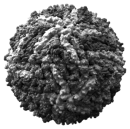
Adenovirus , model of the capsid of a virion
Scheme of an icosahedral virus capsid
Bunyaviridae , scheme of the structure
Schematic cross section through a lambda phage (virus family Siphoviridae )
Scheme of the structure of Arenaviridae
Influenza virus virion
HIV virion
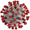
3D graphics of the SARS-CoV-2 virus
Systematic position
Viruses are essentially mere material programs for their own reproduction in the form of a nucleic acid. Although they have specific genetic information, they do not have the synthesis equipment necessary for their replication . Whether viruses can be called living beings depends on the definition of life . A generally accepted, unchallenged definition does not yet exist. Most scientists do not classify viruses as living beings - although the scientific discussion has not yet ended, as, for example, the size of the genome of the cafeteria roenbergensis virus is beginning to be blurred.
Viruses are usually not counted among the parasites because parasites are living things. Some scientists still regard viruses as parasites because they infect a host organism and use its metabolism for their own reproduction. These researchers define viruses as “obligate intracellular parasites” (life forms that are always parasites within a cell) that consist of at least one genome and that require a host cell for replication. Regardless of whether they are classified as living beings or non-living beings, it can be agreed that the behavior of viruses is very similar to that of common parasites. Viruses, like prions , functionless DNA sequences and transposons , can be described as “parasitic” in this sense.
Multiplication and spreading
A virus itself is incapable of any metabolic processes , so it needs host cells to reproduce . The replication cycle of a virus generally begins when a virion attaches itself (adsorption) to a surface protein on a host cell that the virus uses as a receptor . In the case of bacteriophages this is done by injecting its genetic material into a cell; in the case of eukaryotes , the virions are inversed by endocytosis and then penetrate the endosome membrane , e.g. B. by a fusogenic protein . After absorption, a virion must first be freed from its envelopes (uncoating) before replication. The genetic material of the virus, its nucleic acid, is then replicated in the host cell and the envelope proteins and possibly other components of the virions are also synthesized by the host cell using the genes of the virus genome ( protein biosynthesis / gene expression). Thus, in the cell, new viruses are formed ( morphogenesis ), the released as virions, either by the cell membrane is dissolved ( cell lysis , lytic virus replication), or discharged by ( secreted ) are (virus budding, budding) with portions the cell membrane as part of the virus envelope . With the help of immunoevasins , the host's immune defense is suppressed. The number of newly formed virions in an infected host cell is called the burst size .
Another possibility is the integration of the virus genome into that of the host ( provirus ). This is the case with temperate viruses such as the bacteriophage lambda .
The effect of virus replication on the host cell is called the cytopathic effect (CPE). There are different types of cytopathic effect: cell lysis , pycnosis ( poliovirus ), cell fusion ( measles virus , herpes simplex viruses , parainfluenza virus ), intranuclear inclusions ( adenoviruses , measles virus), intraplasmic inclusions ( rabies virus , pox viruses ).
Viruses can be spread in many ways. Viruses pathogenic to humans can be transmitted, for example, via the air by means of droplet infection (e.g. flu viruses) or via contaminated surfaces by smear infection (e.g. herpes simplex ). Plant viruses are often transmitted by insects or by mechanical transmission between two plants, or via contaminated tools in agriculture. An abstract view of the epidemiological kinetics of viruses and other pathogens is developed in theoretical biology .
evolution
origin
The origin of the viruses is not known. Most researchers today assume that viruses are not precursors of cellular life, but rather genes of living things that broke away from living things. Several possibilities are still being discussed, whereby there are basically two different approaches:
- Viruses are very primal; they arose before the first cell and in that chemical “primordial soup” that produced even the most primitive forms of life; along with RNA genomes, they are a holdover from the pre-DNA world. This approach was represented, for example, by Félix Hubert d'Hérelle (1924) and Salvador Edward Luria (1960).
- Viruses are a kind of atrophy of complete organisms that already existed when they were formed.
From this, three theories have been formulated.
- Descent from self-replicating molecules (coevolution). This theory assumes that the origin and evolution of viruses originated from the simplest molecules that were even able to self-duplicate. Then some such molecules would have finally come together to form organizational units that can be viewed as cells. At the same time, other molecules managed to pack themselves into virus particles, which continued to develop in parallel with the cells and became their parasites.
- Virus development through degeneration ( parasite ). This theory is based on the second possible approach presented above, according to which the first viruses originally emerged from free-living organisms such as bacteria (or hypothetical ribocytes ), which slowly and continuously lost more and more of their genetic information until they finally became cell parasites, who rely on a host cell to provide them with the lost functions. A concept that increasing attention in this context (that of Virozelle English virocell ): The actual phenotype of a virus the infected cell, the virion (virus particles), however, only one stage of the reproduction or distribution, similar to pollen or spores .
- Virus formation from the host cell's own RNA or DNA molecules. This third theory, which appears to be the most probable for research, states that viruses arose directly from RNA or DNA molecules in the host cell. As the genetic material of the virus, these nucleic acids that have become independent have acquired the ability to multiply independently of the host cell's genome or its RNA, but in the end they have remained parasites ( S. Luria , 1960). Examples of possible transitional forms are transposons and retrotransposons .
variability
For the evolution of a virus (or any gene), its variability and selection are important. The variability is (as with all organisms) due to copying errors in the replication of the genetic material and serves, among other things, for immune evasion and changing the host spectrum , while selection is often carried out by the host's (immune) response.
More highly organized living beings have developed a very effective possibility of genetic variability through recombination and crossing-over in sexual reproduction, especially in the direction of environmental adaptation and thus further development of their respective species. Virions or viruses, as structures capable of survival , which are dependent on living hosts for their reproduction and thus also spread, without sexual reproduction alone with their ability to mutate show an at least equal opportunity for genetic variability.
It is then ultimately irrelevant that these mutations in the genome of the viruses are basically based on copying errors during replication within the host cells. What counts is the positive effect of the extreme increase in adaptability resulting from this for species conservation. While errors of this kind can lead to cell death in a highly developed mammalian cell , for example , they even have a great selection advantage for viruses .
Copy errors in replication are expressed in point mutations , i.e. in the incorporation of wrong bases at random gene locations . Since viruses, in contrast to the more highly developed cells, have few or no repair mechanisms, these errors are not corrected.
Special forms of genetic change in viruses are described in detail there , for example in the case of influenza viruses, using the terms antigen drift and antigen shift (genetic reassortment ).
Host reactions
Virus infection creates various forms of defense in its hosts . Viruses are only replicated intracellularly because they use the necessary building blocks and enzymes from the cytosol of a host cell for replication . Therefore, various intracellular defense mechanisms have emerged, which are known as restriction or resistance factors . While bacteria including the CRISPR and restriction enzymes to ward off bacteriophages use within a cell, there are eukaryotes such. B. the myxovirus resistance factor Mx1 , the PAMP receptors , the dsRNA-activated inhibitor of translation DAI , the melanoma differentiation antigen 5 ( MDA-5 ), the oligoadenylate synthase OAS1 , the Langerin , the tetherin , the SAM domain and HD domain 1 protein ( SAMHD1 ), the RIG-I , the APOBEC3 , the TRIM5alpha , the protein kinase R and the RNA interference .
In animals, especially vertebrates, an immune response has also developed. It is partly congenital , partly acquired . In the course of the acquired or adaptive immune response, antibodies and cytotoxic T cells are created that can bind individual components of the virus ( antigens ). This enables them to recognize viruses and virus-infected cells and eliminate them.
Coevolution
One observation in the pathogenesis in natural hosts is that pathogens adapted to the host usually do not harm it very much, since they need it for their own development and the immune system is activated by cell damage and apoptosis . Avoiding an immune reaction facilitates replication and transmission (synonymous with transmission ) to other hosts. Some viruses stay in the body for life. Reactivation may occur from time to time, even without symptoms. (See also: Pathogen persistence .) Herpes simplex viruses , for example, reach infection rates (synonymous with contamination ) of over 90% of the German population with less pronounced symptoms. The simian immunodeficiency virus does not produce AIDS in its natural hosts , unlike HIV in humans. In contrast, infections with Ebola virus in humans, but not in their natural hosts, are occasionally self- extinguishing due to their high virulence, before efficient transmission takes place, since the host is severely weakened and soon dies, consequently its range of motion and thus the spread of the virus limited. A severe course of infection with high mortality (see lethality and mortality ) is usually an indication that the causative pathogen has not yet adapted to the organism in question as its reservoir host. However, the transition from pathogens with a high level of replication (and damage caused) to a permanent infection rate ( Infect and persist , avoiding damage) is fluid. In other words, adapted infectious objects tend to persist and a regulated rate of reproduction, while less adapted pathogens tend to lead to premature termination of the chain of infection . Exceptions are e.g. B. H5N1 viruses in birds, Yersinia pestis and human smallpox viruses in humans. However, the adaptation usually takes place on the part of the host, since the pathogens are in competition with their conspecifics and a less reproductive pathogen would perish more quickly. Therefore, a reduction in pathogenicity in pathogens occurs primarily in connection with an increased rate of reproduction.
The adaptation of the host to the pathogen is referred to as host restriction or resistance. The known antiviral and antibacterial mechanisms include, as already explained under host reactions in eukaryotes, in humans, for example, the myxovirus resistance factor Mx1 , the PAMP receptors , the dsRNA-activated translation inhibitor DAI, the MDA5 , the oligoadenylate synthase OAS1 , the langerin , tetherin , APOBEC3 , TRIM5alpha and protein kinase R. In addition, the immune response occurs .
Classification
Conventional virus classification
In 1962, André Lwoff , Robert W. Horne and Paul Tournier introduced a virus taxonomy ("LHT system") based on the binary classification of living things founded by Carl von Linné , which includes the following levels (pattern for the endings of the taxa in Brackets):
- Virosphere ( Phylum : Vira)
This is accompanied by an assignment in groups that are based on the hosts
- Bacteria and archaea (attack by bacteriophages / archaeophages)
- Algae , fungi and protozoa
- Plants (infestation also by viroids )
- Animals, with three subgroups:
- invertebrates (invertebrates)
- Vertebrates (vertebrates)
- Representatives of both groups
Most viruses belong to only one of the above four groups, but virus species in the Rhabdoviridae and Bunyaviridae families can infect both plants and animals. Some viruses only multiply in vertebrates, but are also transmitted mechanically by invertebrates (see vector ), especially by insects . Viruses that rely on the use of genes from other viruses (mamaviruses) during the joint infection of a host cell are called virophages .
Virus taxonomy according to ICTV
The International Committee on Taxonomy of Viruses (ICTV) has developed a classification system to ensure a uniform division into families. The ninth ICTV report defines a concept with the virus species as the lowest taxon in a hierarchical system of branching virus taxa.
Until 2017, the taxonomic structure was basically the same as for the conventional virus classification from level order and below (see above) and was supplemented in 2018 by further levels as follows (with name endings that differ from the LHC system):
- Area (en. Realm) ( ... viria )
- Sub-area (en. Subrealm) ( ... vira ) (ending as with subphylum in the LHC system, as the second top level)
-
Reich (en. Kingdom) ( ... virae )
- Unterreich (en. Subkingdom) ( ... virites )
-
Strain or Phylum ( ... viricota ) (in analogy to ... archaeota - unlike the LHC system, multiple Virusphyla are possible)
- Subphylum ( ... viricotina )
-
Class ( ... viricetes )
- Subclass ( ... viricetidae )
- Order ( ... viral )
- Subordination ( ... virineae )
- Family ( ... viridae )
- Subfamily ( ... virinae )
- Genus or genus ( ... virus )
- Subgenus or subgenus ( ... virus )
- Species or species ( ... virus )
- Subgenus or subgenus ( ... virus )
- Genus or genus ( ... virus )
- Subfamily ( ... virinae )
- Family ( ... viridae )
- Subordination ( ... virineae )
- Order ( ... viral )
- Subclass ( ... viricetidae )
-
Class ( ... viricetes )
- Subphylum ( ... viricotina )
-
Strain or Phylum ( ... viricota ) (in analogy to ... archaeota - unlike the LHC system, multiple Virusphyla are possible)
- Unterreich (en. Subkingdom) ( ... virites )
-
Reich (en. Kingdom) ( ... virae )
- Sub-area (en. Subrealm) ( ... vira ) (ending as with subphylum in the LHC system, as the second top level)
Of these permitted levels so far (ICTV as of February 2019) only area, phylum, subphylum, class, order, subordination, family, subfamily, genus, subgenus and species are in actual use. There is no definition of subspecies , strains (in the sense of varieties, such as “bacterial strain”) or isolates in these guidelines . The name endings of all ranks thus have “vir” as a component (but not in the form “viroid”); the abbreviations end with "V", possibly followed by a number (not Roman, but Arabic). For viroids and satellites as subviral particles , an analogous taxonomy can be used, each with its own name endings with a characterizing component.
As of March 2019, the following regulations have been added:
To the Phylum Negarnaviricota :
Further orders and subordinates of the Riboviria :
Orders not grouped into higher ranks:
The Baltimore Classification

The classification proposed by Nobel Laureate and biologist David Baltimore is based on the exact form in which the virus genome is and how the messenger RNA ( mRNA ) is generated from it. The virus genome can be in the form of DNA or RNA, single strand (English: single-stranded, ss) or double strand (English double-stranded, ds). A single strand can exist as an original (English: sense, +) or in a complementary form (English: antisense, -). Under certain circumstances, an RNA genome is temporarily converted into DNA for replication ( retroviruses ) or, conversely, a DNA genome is temporarily transcribed into RNA ( pararetroviruses ); in both cases the RNA is written back into DNA with a reverse transcriptase (RT).
The entire virosphere is defined by the following seven groups:
- I: dsDNA viruses (also adenoviruses , herpes viruses , giant viruses , smallpox viruses )
- II: ssDNA viruses (+ strand) DNA (plus parvoviruses )
- III: dsRNA viruses (also reoviruses )
- IV: (+) ssRNA viruses (+ strand) RNA (also picornaviruses , togaviruses )
- V: (-) ssRNA viruses (- strand) RNA (also orthomyxoviruses , rhabdoviruses )
- VI: ssRNA-RT viruses (+ strand) - RNA with DNA intermediate stage ( retroviruses )
- VII: dsDNA-RT viruses - DNA with RNA intermediate stage ( pararetroviruses , plus hepadnaviruses )
Modern virus classifications use a combination of ICTV and Baltimore.
Spelling of the virus type names
The official international, scientific name of a virus is the English-language name, which is also used in the international abbreviation, such as Lagos bat virus (LBV). This abbreviation is also used unchanged in German. Logically, the abbreviation for the German virus name Lagos-Fledermaus-Virus is also LBV .
In English virus names, such as West Nile virus , no hyphens are normally used and virus is lowercase. The hyphen appears in English only with adjectives , i.e. with Tick-borne encephalitis virus or Avian encephalomyelitis-like virus .
In German the virus name be partially written with hyphens , that West Nile virus , hepatitis C virus , human herpes virus , Lagos bat virus , European bat Lyssa virus , sometimes together. In front of the numbers of subtypes (as in English) there is a space , for the abbreviations a hyphen, e.g. B. European Bat Lyssa Virus 1 (EBLV-1), Herpes Simplex Virus 1 (HSV-1) and Human Herpes Virus 1 (HHV-1).
Viruses pathogenic to humans and diseases caused
Viruses can cause a wide variety of diseases in humans. But these human pathogenic viruses are here in terms genome and Behüllung classified and their taxonomy by ICTV listed.
Enveloped viruses
Double-stranded DNA viruses = dsDNA
- Family Poxviridae
- Subfamily Chordopoxvirinae
- Genus Orthopoxvirus
- Orthopoxvirus variola = Variolavirus - smallpox , real smallpox
- Orthopoxvirus variola var. Alastrim = Kaffirpoxvirus - smallpox , white smallpox
- Monkeypoxvirus (MPV) also Orthopoxvirus simiae ; Monkey pox virus - monkey pox ; also transferable to humans, symptoms as with human pox, but much milder
- Genus Parapoxvirus
- Parapoxvirus ovis = Orf virus - Orf
- Genus Molluscipoxvirus
- Molluscum Contagiosum Virus - Dellwarze ( Molluscum contagiosum )
- Genus Orthopoxvirus
- Subfamily Chordopoxvirinae
- Family Herpesviridae
- Subfamily Alphaherpesvirinae
- Genus simplex virus
- Herpes simplex virus 1 (HSV-1) = Human herpes virus 1 (HHV-1) - herpes simplex , herpes labialis , aphthous stomatitis
- Herpes Simplex Virus 2 (HSV-2) = Human Herpes Virus 2 (HHV-2) - Herpes simplex , genital herpes
- Herpes B virus = ( Herpes virus simiae )
- Genus Varicellovirus
- Varicella zoster virus (VZV) = human herpes virus 3 (HHV-3) - chickenpox = varicella (herpes zoster), shingles
- suid herpesvirus type 1 (SHV-1) = pseudowut virus , Aujeszky virus and the like a. - Aujeszky's disease = pseudo rage, itch epidemic, mad scabies, etc. a. (in animals, with low pathogenicity also transferable to humans)
- Genus simplex virus
- Subfamily Betaherpesvirinae
- Genus Cytomegalovirus
- Human Cytomegalovirus (HCMV) = Human Cytomegalovirus (HZMV) = Human Herpes Virus 5 (HHV-5) - Cytomegaly
- Genus Reseolovirus
- Human herpes virus 6 (HHV-6) - three-day fever
- Human herpes virus 7 (HHV-7) - three day fever
- Genus Cytomegalovirus
- Subfamily Gammaherpesvirinae
- Genus Lymphocryptovirus
- Epstein Barr Virus (EBV) = Human Herpes Virus 4 (HHV-4) - Pfeiffer glandular fever , Burkitt's lymphoma
- Genus Rhadinovirus
- Human Herpes Virus 8 (HHV-8) - Kaposi's sarcoma
- Genus Lymphocryptovirus
- Subfamily Alphaherpesvirinae
- Family Hepadnaviridae
- Genus Orthohepadnavirus
- Hepatitis B Virus (HBV) - Hepatitis B
- Genus Orthohepadnavirus
Single (+) strand RNA viruses = ss (+) RNA
- Togaviridae family
- Genus Alphavirus - causative agent of arboviruses
- Barmah Forest Virus - Barmah Forest fever with flu-like symptoms, epidemic polyarthritis
- Chikungunya virus (CHIKV) - Chikungunya fever
- Eastern Equine Encephalitis Virus (EEEV) = Eastern
- Genus Alphavirus - causative agent of arboviruses
- Western Equine Encephalitis Virus (WEEV) = Western equine encephalitis virus - transmission by mosquitoes to humans is also possible (rare!) → Western equine encephalomyelitis ( encephalitis / encephalomyelitis )
- Everglades Virus - Everglades Fever
- O'nyong Nyong Virus (ONNV) - O'nyong Nyong Fever
- Mayaro fever virus (MAYV) - Mayaro fever
- Semliki Forest Virus (SFV) - Semliki Forest fever
- Mucambo Virus - Mucambo Fever
- Ross River Virus (RRV) - Ross River Fever
- Sindbis virus (SINV) - Sindbis fever (inflammation of the joints ["epidemic polyarthritis "], sometimes with rashes and rarely with encephalitis )
- Genus Hepacivirus
- Hepatitis C Virus (HCV) - Hepatitis C
- GB-Virus-C (no disease value)
- Genus Flavivirus
- West Nile Virus (WNV) - West Nile fever
- Dengue virus (DENV) - Dengue fever
- Yellow fever virus (YFV) - yellow fever
- Louping Ill Virus (LIV) - Louping Ill Encephalitis
- St. Louis Encephalitis Virus (SLEV) - St. Louis Encephalitis
- Japan Encephalitis Virus (JEV) - Japanese encephalitis
- Usutu virus (USUV) - non-specific symptoms such as fever and / or rashes
- Kyasanur Forest Disease Virus (KFDV) - Kyasanur Forest Fever
- Powassan virus (POWV) - Powassan encephalitis
-
TBE virus [English: tick-borne encephalitic virus (TBEV)] - TBE (early summer meningoencephalitis)
- Subtype European / Western tick-borne encephalitis virus (WTBEV)
- Subtype Siberian tick-borne encephalitis virus (STBEV)
- Subtype Far-Eastern tick-borne encephalitis virus (Far-Eastern TBEV); formerly Russian Spring Summer Encephalitis Virus (RSSEV) - RSSE , also RFSE (Russian Spring Summer Encephalitis , Russian Early Summer Encephalitis )
- Zika virus (ZIKV) (2 main groups; various subtypes) - mostly just skin rash, fever, joint pain, conjunctivitis
- Subfamily Orthocoronavirinae
- Genus Alphacoronavirus
- Subgenus Duvinacovirus
- Human coronavirus 229E (HCoV-229E) - common cold
- Setracovirus subgenus
- Human coronavirus NL63 (HCoV-NL63) - common cold
- Subgenus Duvinacovirus
- Genus Betacoronavirus
- Subgenus Embecovirus
- Species betacoronavirus 1
- Subspecies human coronavirus OC43 (HCoV-OC43) - common cold; occasionally severe respiratory tract infection, pneumonia
- Species human coronavirus HKU1 (HCoV-HKU1) - common cold
- Species betacoronavirus 1
- Subgenus Merbecovirus
- Middle East respiratory syndrome coronavirus (MERS-CoV) - flu-like symptoms, severe respiratory infection, pneumonia and possibly kidney failure
- Subgenus Sarbecovirus
-
SARS-associated coronavirus (SARS-CoV) - SARS ( atypical pneumonia ), with subtype
- Subtype SARS-CoV-2 (eng. 2019-novel Coronavirus, 2019-nCoV, or Wuhan seafood market pneumonia virus) - COVID-19 : Infection of the lower respiratory tract up to pneumonia
-
SARS-associated coronavirus (SARS-CoV) - SARS ( atypical pneumonia ), with subtype
- Subgenus Embecovirus
- Genus Alphacoronavirus
- Subfamily Torovirinae
- Genus Torovirus
- various types - gastroenteritis
- Genus Torovirus
- Subfamily Orthoretrovirinae
- Genus Deltaretrovirus
- Human T-Lymphotropic Virus 1 (HTLV-1) - Adult T-Cell Leukemia , Tropical Spastic Paraparesis
- Human T-Lymphotropic Virus 2 (HTLV-2) - Leukemia (?)
- Human T-lymphotropic virus 3 (HTLV-3) - unknown
- Human T Lymphotropic Virus 4 (HTLV-4) - unknown
- Genus Lentivirus
- Human Immunodeficiency Virus Type 1 (HIV-1) - AIDS
- Human Immunodeficiency Virus Type 2 (HIV-2) - AIDS
- Genus Deltaretrovirus
Single (-) - strand RNA viruses = ss (-) RNA
- Family Arenaviridae
- Genus Arenavirus
- Chapare virus - hemorrhagic fever
- Lassa virus - Lassa fever
- lymphocytic chorio-meningitis virus (LCMV) - lymphocytic choriomeningitis
- Lujo Virus - Hemorrhagic Fever
- Tacaribe Virus - Hemorrhagic Fever
- Junin virus - Junin fever (Argentine hemorrhagic fever )
- Machupo virus - Machupo fever (Bolivian hemorrhagic fever )
- Genus Arenavirus
- Family Bornaviridae
- Genus Bornavirus
- Virus of Borna disease (English Borna disease virus = BoDV.) - causative agent of Borna disease in horses and sheep, and perhaps also applicable to humans - Mood Disorders
-
Mammalian Bornavirus 1
- Red squirrel bornavirus 1 ( Variegated squirrel Bornavirus 1 = VSBV-1 ) detected in red squirrels ( Sciurus variegatoides ), also transmissible to humans - potentially fatal encephalitis
- Genus Bornavirus
- Family Bunyaviridae - causative agents of arboviruses
- Genus Orthobunyavirus
- Bunyamwera virus (serogroup)
- Batai virus (BATV) - flu-like symptoms and rashes
- California Encephalitis Virus (Serogroup) - Encephalitis
- Genus phlebovirus
- Rift Valley Fever Virus (3 subtypes) - Rift Valley Fever
-
Sandfly fever virus (SFNV) - Sandfly fever = sandfly fever
- Subtype Karimabad virus (KARV)
- Subtype sand fly fever virus Sabin (SFNV-Sabin)
- Subtype Tehran virus (THEV)
- Subtype Toscana virus (TOSV) - Pappataci fever
- Serotypes : Tuscany (T), Sicily (S) and Naples (N)
- Genus Nairovirus
-
Crimean Congo Fever Virus (serogroup):
- Subtype Crimean Congo Hemorrhagic Fever Virus (CCHFV) - Crimean Congo fever
- Hazara virus (HAZV) subtype - Crimean-Congo fever
- Subtype Khasan virus (KHAV) - Crimean-Congo fever
-
Crimean Congo Fever Virus (serogroup):
- Genus Hantavirus
- Hantaan virus (4 subtypes) - hemorrhagic fever , nephritis
- Seoul virus (serogroup) - hemorrhagic fever
- Prospect Hill virus (2 subtypes) - hemorrhagic fever
- Puumala virus (serogroup) - hemorrhagic fever , pneumonia , nephritis
- Dobrava Belgrade Virus - Hemorrhagic Fever
- Tula virus - hemorrhagic fever
- Sin Nombre Virus (serogroup) - hemorrhagic fever with severe pulmonary edema
- Genus Orthobunyavirus
- Filoviridae family
- Genus Marburg virus
- Lake Victoria Marburg virus (serogroup) - Marburg fever ( hemorrhagic fever )
- Genus Ebola virus
- Zaire Ebolavirus (ZEBOV) serogroup - Ebola fever ( hemorrhagic fever )
- Sudan Ebolavirus (SEBOV) serogroup - Ebola fever ( hemorrhagic fever )
- Reston Ebolavirus (REBOV) serogroup - not pathogenic to humans, only
- Genus Marburg virus
- Côte d'Ivoire Ebolavirus (CIEBOV) serogroup - Ebola fever ( hemorrhagic fever )
- Bundibugyo Ebola virus (BEBOV) serogroup - Ebola fever ( hemorrhagic fever )
- Genus Influenzavirus A - Influenza (flu)
- Influenza virus A variant H1N1 - Influenza (flu)
- Influenza virus A variant H3N2 - influenza (flu)
- (Avian) influenza virus A variant H5N1 , highly pathogenic avian influenza virus ( HPAIV ) - "bird flu", in animals, also transmissible to humans, but hardly from person to person.
- Genus Influenzavirus B - Influenza (flu)
- Influenza Virus B / Victoria Line - Influenza (Flu)
- Influenza Virus B / Yamagata Line - Influenza (Flu)
- Genus Influenzavirus C - Influenza (flu)
- Genus Influenzavirus D - Influenza (flu)
- Genus Avulavirus
- Human parainfluenza virus (types 1, 3) - common cold , parainfluenza
- Genus Morbillivirus
- Genus Henipavirus
- Hendra Virus , (formerly Equines Morbillivirus ) - Pneumonia ; Encephalitis
- Nipah Virus - Pneumonia ; Encephalitis
- Genus Rubula Viruses
- Human parainfluenza virus (type 2, 4) - common cold , parainfluenza
- Mumps virus - mumps
- Genus Orthopneumovirus (formerly: Pneumovirus )
- Human Respiratory Syncytial Virus (HRSV) (Type A, B) - respiratory tract infection , common cold
- Genus metapneumovirus
- Human metapneumovirus (HMPV) (types A1 to 2, B1 to 2) - respiratory infection , common cold
- Genus Vesiculovirus
- Vesicular stomatitis Indiana virus (VSV) - stomatitis vesicularis (inflammation of the oral mucosa with blistering) in animals, also transmitted to humans
- Genus Lyssavirus
- Rabies virus (RABV) (formerly genotype 1) = rabies virus - rabies , in animals, can also be transmitted to humans
- Mokola virus (MOKV) (formerly genotype 3) - rabies , in animals, can also be transmitted to humans
- Duvenhage virus (DUVV) (formerly genotype 4) - rabies , in animals, can also be transmitted to humans
- European bat lyssa virus 1 + 2 (EBLV-1, -2) (formerly genotypes 5 and 6) - rabies , in animals, also transmitted to humans
- Australian Bat Lyssa Virus (ABLV) (formerly genotype 7) - rabies , in animals, also transmitted to humans
Unenveloped viruses
Double-stranded DNA viruses = dsDNA
- Family Adenoviridae
- Genus mastadenovirus
- Human adenovirus AF (51 subtypes) - runny nose , colds , diarrhea
- Genus mastadenovirus
- Family Polyomaviridae
- Genus polyomavirus
- BK polyomavirus (BKPyV) = BK virus (BKV) = polyoma virus hominis type 1 - results in immunosuppressive treatment after transplantation possibly the loss of the. Graft
- JC polyomavirus (JCPyV) = JC virus (JCV) = polyomavirus hominis type 2 - in cellular immunosuppressed ( AIDS ) to progressive multifocal leukoencephalopathy (PML)
- Genus polyomavirus
- Family Papillomaviridae
- Genus papillomavirus
- Subgenus human papillomavirus
- various human papilloma viruses (HPV) - warts
- Condyloma Virus 6 (HPV-6) - genital warts
- Condyloma Virus 11 (HPV-11) - genital warts
- Human papillomavirus 16/18/30… (HPV-16 / -18 / -30…) - cervical cancer (cervical cancer)
- Subgenus human papillomavirus
- Genus papillomavirus
Single stranded DNA viruses = ssDNA
- Family Parvoviridae
-
- Subfamily Parvovirinae
-
- Genus Dependoparvovirus (aka Dependovirus )
-
- Species adeno-associated virus A (AAV-A)
- Adeno-associated virus 1 to 4 (AAV-1 to AAV-4)
- Species adeno-associated virus B (AAV-B)
- Adeno-associated virus 5 (AAV-5)
- Genus Erythroparvovirus (aka Erythrovirus )
-
- Species Primate erythroparvovirus 1
Double-stranded RNA viruses = dsRNA
- Reoviridae family
- Genus Rotavirus
- various types - gastroenteritis with diarrhea
- Genus Coltivirus
- Genus Rotavirus
Single (+) strand RNA viruses = ss (+) RNA
- Family Caliciviridae
- Genus norovirus
-
Norovirus (NV) = Norwalk-Like-Virus (NLV)
- Human noroviruses of groups GGI, GGII and GGIV - vomiting diarrhea = gastroenteritis
-
Norovirus (NV) = Norwalk-Like-Virus (NLV)
- Genus sapovirus
- Sapovirus (SV) - gastroenteritis
- Genus norovirus
- Family Hepeviridae
- Genus Hepevirus
- Hepatitis E Virus (HEV) - Hepatitis E.
- Genus Hepevirus
- Family Picornaviridae
- Enterovirus genus
- Poliovirus type 1-3 - polio
-
Coxsackievirus A / B - from the common cold to meningitis , pancreatitis or myocarditis , rarely paralysis
- Coxsackievirus B1 (CVB-1) to B 6 - common cold
- Echovirus - exanthema enanthemum , infections of the upper respiratory tract ( cold ), herpangina , myopericarditis , disseminated (disseminated) infection in newborns, chronic meningoencephalitis in immunocompromised patients, meningitis , encephalitis rarely paralysis
-
Enterovirus
-
Human enteroviruses - common cold
- Human Enterovirus 70 (EV-70) - acute hemorrhagic conjunctivitis
- Human Enterovirus 71 (EV-71) - meningoencephalitis , rash and poliomyelitis like syndrome = hand-foot-mouth disease
-
Human enteroviruses - common cold
- Genus hepatovirus
- Genus rhinovirus
-
Rhinovirus
- Human rhinoviruses -1 A (HRV-1 A) or 1 B to 100 - common cold
-
Rhinovirus
- Enterovirus genus
Oncoviruses
The group of " oncoviruses ", the most important carcinogenic viruses in humans, is responsible for 10 to 15 percent of all human cancers worldwide , according to estimates by the American Cancer Society even for around 17% of cancer cases.
- Epstein Barr Virus (EBV)
- Hepatitis B virus (HBV)
- Hepatitis C virus (HCV)
- Human papillomavirus (HPV)
- Human T Lymphotropic Virus 1 (HTLV-1)
- Human herpesvirus 8 (HHV-8, also Kaposi's sarcoma herpesvirus, KSHV)
Giant viruses
Antiviral drugs
Since viruses or virions, unlike bacteria, are not cells, they cannot be killed like such. It is only possible to prevent or prevent a viral infection and virus replication using antivirals . In particular, the biochemical reproduction processes can be very different from virus type to virus type, which makes it difficult to find an inhibiting or suppressing agent.
Since the virus replicates inside normal cells and there it is very closely linked to the central biochemical cell mechanisms, the antiviral agents in question must either
- prevent virions from entering the host cells,
- intervene in the cell metabolism to the detriment of virus replication
- or stop the escape of the new viruses from the cells after a possible virus replication in the cells.
On the other hand, these sought-after active ingredients must also be compatible with the body metabolism, the cell structure and / or the internal cell metabolism as a whole, as otherwise not only, for example, virus replication in the cells comes to a standstill, but in the worst case also the (cell) life of the entire organism being treated .
Because these conditions are very difficult to reconcile, the antiviral drugs developed so far are often associated with serious side effects . This balancing act posed difficult tasks for medicine, which so far have mostly remained unsolved.
The development of effective antiviral drugs is also exacerbated by the development of resistance on the part of the viruses to be combated to a useful active ingredient once found, of which they are well capable due to their extremely fast reproduction cycle and the biochemical nature of this replication.
Virus therapy
There is currently more research into therapies that use viruses to heal diseases. This research includes the use of viral vectors as oncolytic viruses to combat tumors , as phage therapy for the targeted infection and lysis of bacteria , some of which are resistant to antibiotics , as a vaccine for the prophylaxis and therapy of infectious diseases , for the generation of induced pluripotent stem cells or for gene therapy of genetic defects .
See also
literature
- Older literature
- Feodor Lynen : The Virus Problem. In: Angewandte Chemie Vol. 51. No. 13, 1938, ISSN 0044-8249 , pp. 181-185.
- Current literature
- Hans W. Doerr, Wolfram H. Gerlich (eds.): Medical virology - basics, diagnosis and therapy of virological diseases. Thieme, Stuttgart / New York 2002, ISBN 3-13-113961-7 .
- Walter Doerfler: Viruses. Fischer Taschenbuch Verlag, Frankfurt a. M. 2002, ISBN 3-596-15369-7 .
- Dietrich Falke , Jürgen Bohl u. a .: bedside virology: clinic, diagnostics, therapy. Springer, Heidelberg et al. 1998, ISBN 3-540-64261-7 . (with references)
- Dietrich Falke, Jürgen Podlech: Viruses. In: Peter Reuter: Springer Lexicon Medicine. Springer, Berlin a. a. 2004, ISBN 3-540-20412-1 , pp. 2273-2282.
- SJ Flint, LW Enquist, VR Racaniello (Eds.): Principles of Virology. 2nd edition, ASM Press, Washington DC 2003, ISBN 1-55581-259-7 .
- Alfred Grafe: Viruses - parasites of our living space. Springer, Berlin / Heidelberg / New York 1977, ISBN 3-540-08482-7 .
- David M. Knipe, Peter M. Howley, et al. (Ed.): Fields' Virology. 2 volumes, 5th edition, Lippincott Williams & Wilkins, Philadelphia 2007, ISBN 978-0-7817-6060-7 (standard work in virology).
- Arnold J. Levine : Viruses: Thieves, Murderers, and Pirates. Spektrum Akademischer Verlag, Heidelberg 1992, ISBN 3-86025-073-6 .
- Susanne Modrow, Dietrich Falke , Uwe Truyen: Molecular Virology. An introduction for biologists and medical professionals. (= Spectrum textbook ). 2nd edition, Spektrum Akademischer Verlag, Heidelberg 2002, ISBN 3-8274-1086-X .
- Stephen S. Morse: The Evolutionary Biology of Viruses. Raven Press, New York 1994, ISBN 0-7817-0119-8 .
- Sven P. Thoms: Origin of life: how and when did life arise on earth? ... (= Fischer pocket books; Fischer compact. ). Fischer Taschenbuch Verlag, Frankfurt a. M. 2005, ISBN 3-596-16128-2 .
- Luis P. Villarreal: Viruses and the Evolution of Life. ASM Press, Washington 2005, ISBN 978-1-55581-309-3 .
- Ernst-Ludwig Winnacker : Viruses: The secret rulers. How flu, AIDS and hepatitis threaten our world. Eichborn, Frankfurt a. M. 1999, ISBN 3-8218-1598-1 .
- Gottfried Schuster: Viruses in the Environment. Teubner, Stuttgart 1998, ISBN 3-519-00209-4 .
- Dorothy H. Crawford: The invisible enemy: a natural history of viruses. Oxford Univ. Press, Oxford 2002, ISBN 0-19-856481-3 .
- Brian W. Mahy: The dictionary of virology. Elsevier, Amsterdam 2008, ISBN 0-12-373732-X .
- Susanne Modrow: Viruses: Basics, Diseases, Therapies. [An easily understandable introduction for medical laypeople]. Beck, Munich 2001. ISBN 3-406-44777-5
- Hartwig Klinker: Virus infections. In: Marianne Abele-Horn (Ed.): Antimicrobial Therapy. Decision support for the treatment and prophylaxis of infectious diseases. With the collaboration of Werner Heinz, Hartwig Klinker, Johann Schurz and August Stich, 2nd, revised and expanded edition. Peter Wiehl, Marburg 2009, ISBN 978-3-927219-14-4 , pp. 297-307.
- Marilyn J. Roossinck: Viruses! Helpers, enemies, bon vivants - in 101 portraits . Springer, Berlin 2018, ISBN 978-3-662-57543-7 .
- Sunit K. Singh (Ed.): Viral Infections and Global Change. [On the influence of globalization and climate change on the spread and transmission of viruses, especially tropical viruses]. Wiley Blackwell, Hoboken NJ 2014, ISBN 978-1-118-29787-2 (print); ISBN 978-1-118-29809-1 (eBook).
Web links
- Viruses: structure, specific characteristics, development, cell biology, differentiation from bacteria
- The Universal Virus Database (English)
- ViralZone , Swiss Institute of Bioinformatics (SIB) - database with all known viruses (English)
- HowStuffWorks.com: How Viruses Work (English)
- Virusworld ( 3-D representations of viruses derived from X-ray structure analyzes )
- How viruses spurred human evolution , riffreporter
Individual evidence
- ^ Karl Ernst Georges : Comprehensive Latin-German concise dictionary . 8th, improved and increased edition. Hahnsche Buchhandlung, Hannover 1918 ( zeno.org [accessed on January 21, 2020]).
- ↑ Duden online: Virus, that or that
- ↑ Karin Mölling: Superpower of Life. Travel to the amazing world of viruses. 1st edition, Beck, Munich 2015, ISBN 978-3-406-66969-9 .
- ↑ Tens of thousands of unknown viruses in wastewater . On: scinexx.de of October 6, 2011, last accessed on September 17, 2014.
- ^ Paul G. Cantalupo et al .: Raw Sewage Harbors Diverse Viral Populations. In: mBio Vol. 2, No. 5, e00180-11, doi: 10.1128 / mBio.00180-11 ( full text as PDF file ).
- ↑ TA McAllister et al .: "Ruminant Nutrition Symposium: Use of genomics and transcriptomics to identify strategies to lower ruminal methanogenesis", in: ACSESS DL, 2015 Archived copy ( Memento from April 7, 2016 in the Internet Archive ) doi: 10.2527 / jas .2014-8329
- ↑ Shmoop Biology: Phages Shmoop University, 2016
- ↑ Pierer's Universal Lexicon of the Past and Present . 4th edition. Publishing house by HA Pierer , Altenburg 1865 ( zeno.org [accessed on January 21, 2020] encyclopedia entry "Virus").
- ^ Death of the Rev. Dr. Peckwell In: The Times, Aug 23, 1787, p. 2.
- ↑ Florian Witzmann et al .: Paget disease of bone in a Jurassic dinosaur. In: Current Biology. Volume 21, No. 17, R647-R648, 2011, doi: 10.1016 / j.cub.2011.08.006 ( full text as PDF file ).
- ↑ John B Carter, Venetia A Saunders: Virology: Principles and Applications. 1st edition, Wiley, Chichester UK 2007, ISBN 0-470-02387-2 , p. 6.
- ^ Matthias G. Fischer, Michael J. Allen, William H. Wilson, and Curtis A. Suttle: Giant virus with a remarkable complement of genes infects marine zooplankton . In: Proceedings of the National Academy of Sciences . 2010. doi : 10.1073 / pnas.1007615107 .
- ^ Salvador Edward Luria, James E. Darnell : General Virology. 3rd edition, John Wiley & Sons, New York et al. a. 1978, ISBN 978-0-471-55640-4 .
- ↑ Luis P. Villarreal, Guenther Witzany: Viruses are essential agents within the roots and stem of the tree of life. In: Journal of Theoretical Biology. Vol. 262, No. 4, 2010, pp. 698-710, doi: 10.1016 / j.jtbi.2009.10.014 .
- ↑ P. Forterre: The virocell concept and environmental microbiology . In: The ISME Journal . 7, 2012, pp. 233-236. doi : 10.1038 / ismej.2012.110 . PMC 3554396 (free full text).
- ↑ David M. Needham, Susumu Yoshizawa, Toshiaki Hosaka, Camille Poirier, Chang Jae Choi, Elisabeth Hehenberger, Nicholas AT Irwin, Susanne Wilken, Cheuk-Man Yung, Charles Bachy, Rika Kurihara, Yu Nakajima, Keiichi Kojima, Tomomi Kimura-Someya , Guy Leonard, Rex R. Malmstrom, Daniel R. Mende, Daniel K. Olson, Yuki Sudo, Sebastian Sudek, Thomas A. Richards, Edward F. DeLong, Patrick J. Keeling, Alyson E. Santoro, Mikako Shirouzu, Wataru Iwasaki , Alexandra Z. Worden: A distinct lineage of giant viruses brings a rhodopsin photosystem to unicellular marine predators , in: PNAS, 23 September 2019, doi: 10.1073 / pnas.1907517116 , ISSN 0027-8424, PDF
- ↑ Georg Löffler, Petro E Petrides (Ed.): Biochemie und Pathobiochemie (= Springer textbook. ) 7th, completely revised edition, Springer, Berlin / Heidelberg / New York 2003, ISBN 3-540-42295-1 .
- ↑ David Moreira, Purificación López-García: Ten reasons to exclude viruses from the tree of life. In: Nature Reviews Microbiology . Vol. 7, April 2009, pp. 306-311, doi: 10.1038 / nrmicro2108 .
- ↑ a b V. J. Torres, DL Stauff et al .: A Staphylococcus aureus regulatory system that responds to host heme and modulates virulence. In: Cell host & microbe. April 19, 2007, Volume 1, No. 2, pp. 109-19, PMID 18005689 , PMC 2083280 (free full text).
- ↑ G. Silvestri: Naturally SIV-infected sooty mabeys: are we closer to understanding why they do not develop AIDS? In: Journal of Medical Primatology. (J Med Primatol.) 2005, Vol. 34, No. 5-6, pp. 243-52, PMID 16128919 .
- ↑ MJ Pantin-Jackwood, DE Swayne: Pathogenesis and pathobiology of avian influenza virus infection in birds. In: Revue scientifique et technique (International Office of Epizootics). (Rev Sci Tech.) 2009, Vol. 28, No. 1, pp. 113-36, PMID 19618622 .
- ↑ KD Mir, MA Gasper, V. Sundaravaradan, DL Sodora: SIV infection in natural hosts: resolution of immune activation during the acute-to-chronic transition phase. In: Microbes and Infection. 2011, Volume 13, No. 1, pp. 14-24, PMID 20951225 , PMC 3022004 (free full text).
- ↑ MJ. Adams, EJ. Lefkowitz, AM. King, EB. Carstens: Recently agreed changes to the International Code of Virus Classification and Nomenclature . In: Archives of Virology . 158, No. 12, December 2013, pp. 2633-9. doi : 10.1007 / s00705-013-1749-9 . PMID 23836393 .
- ↑ International Committee on Taxonomy of Viruses Executive Committe , Virus Taxonomy: 2018 Release , How to write virus and species names
- ↑ International Committee on Taxonomy of Viruses Executive Committee : The new scope of virus taxonomy: partitioning the virosphere into 15 hierarchical ranks , in: Nature Microbiology Volume 5, pp. 668-674 of April 27, 2020, doi: 10.1038 / s41564-020 -0709-x ; and Nadja Podbregar: A family tree for the virosphere , on: scinexx.de from April 29, 2020. Both articles have the status of January 2020, i. H. Details of the Master Species List (MSL) No. 35 of the ICTV from March 2020 have not yet been taken into account. However, this has no meaning for the basic intention of ICTV, with MSL # 35 the development has only continued in the specified direction.
- ↑ International Committee on Taxonomy of Viruses (ICTV): ICTV Master Species List 2018b.v2 (MSL # 34v)
- ^ Susanne Modrow, Dietrich Falke, Uwe Truyen: Molecular Virology. 2nd edition, Spektrum - Akademischer Verlag, Heidelberg / Berlin 2003, ISBN 3-8274-1086-X .
- ↑ Th. Mertens, O. Haller, H.-D. Klenk (Hrsg.): Diagnosis and therapy of viral diseases - guidelines of the society for virology. 2nd edition, Elsevier / Urban & Fischer, Munich 2004, ISBN 3-437-21971-5 .
- ↑ Thomas Berg, Norbert Suttorp: Infectious Diseases. Thieme, Stuttgart 2004, ISBN 3-13-131691-8 .
- ↑ Lexicon of Medical Laboratory Diagnostics . Pp. 402-403: Barmah Forest Viruses (BFV).
- ↑ Gerhard Dobler, Horst Aspöck: Arboviruses transmitted by mosquitoes as causative agents of human infections. In: Horst Aspöck (ed.): Sick through arthropods (= Denisia. Volume 30). Biology Center (Upper Austrian State Museums in Linz), 2010, ISSN 1608-8700 , here p. 518: Barmah Forest Fever. - 2.6.4. Clinic ( full text as PDF ).
- ^ Centers for Disease Control and Prevention (CDC): Eastern Equine Encephalitis . On: cdc.gov of April 5, 2016; last accessed on August 30, 2016.
- ↑ Adriana Delfraro, Analía Burgueño, Noelia Morel u. a .: Fatal Human Case of Western Equine Encephalitis, Uruguay. In: Emerging Infectious Diseases. Volume 17, No. 5, May 2011, pp. 952–954 → Letters, doi: 10.3201 / eid1705.101068 ( full text as PDF file ).
- ↑ Bernd Hoffmann, Dennis Tappe, Dirk Höpe and others: A Variegated Squirrel Bornavirus Associated with Fatal Human Encephalitis. In: The New England Journal of Medicine. (N Engl J Med) 2015, Volume 373, pp. 154-16, doi: 10.1056 / NEJMoa1415627 .
- ↑ NCBI: Dependovirus (genus)
- ↑ D. Martin, JS Gutkind: Human tumor-associated viruses and new insights into the molecular mechanisms of cancer . In: Oncogene . Vol. 27, No. 2, 2008, pp. 31-42. PMID 19956178 .
- ↑ C. Zimmer: Cancer - a side effect of evolution? In: Spectrum of Science. No. 9, 2007, pp. 80-88.
- ^ M. Stadtfeld et al .: Induced Pluripotent Stem Cells Generated Without Viral Integration. On: science-online of September 25, 2008, doi: 10.1126 / science.1162494 .


